Kansas Chronic Wasting Disease CWD TSE Prion Update 143 Confirmed Cases To Date
Kansas Chronic Wasting Disease CWD TSE Prion
The first case of CWD was found in a captive bull elk in Harper County in 2001.
As of 30 June 2017, CWD has been detected in 143 wild, free-ranging cervids in Deer Management Units 1, 2, 3, 4, 5, 7,17, 18 – 1 captive elk, 11 mule deer, and 131 white-tailed deer.
Surveillance efforts began in 1996.
Hunters and other wildlife enthusiasts can avoid the human-assisted spread of CWD by not transporting a live or dead deer or elk from areas where CWD occurs to CWD-free areas.
There is currently no known treatment or eradication method for CWD, so preventing the introduction of the the disease into new areas is of utmost importance to the health of local deer herds.
Baiting and feeding deer tend to concentrate deer at small point on the landscape, often with the trails leading to the feeding sites resembling the wheel spokes of a bicycle.
Anytime animals are concentrated at this type of "hub," the likelihood of disease transmission increases in a deer herd. More alarming, the transferring of CWD prions to healthy deer is not the only concern. Diseases such as bovine tuberculosis, foot rot, and fungal infections; and a host of detrimental parasites, including exotic lice, flukes, mange mites, lungworms, and barberpole worms are transmitted more efficiently when deer are concentrated in a small area, especially around feeding stations.
From the 2015-2016 sample, prevalence was calculated to be between 10-20% with 95% confidence in bucks 2.5 years-old and older in the Northwest Zone.
Another major concern is the potential for spread of CWD from captive cervid farms into the wild cervid population. Once a disease gets into a wild population, it is virtually impossible eradicate. The only thing that can be done is control the spread of the disease at great expense. KDWPT recommends that every captive cervid operator enroll in the voluntary CWD monitoring program administered by the Kansas Department of Agriculture's Kansas Animal Health Division. The sooner diseases such as CWD can be detected in captives, the sooner control efforts can begin and possibly prevent the spread of disease to wild populations of the state. CWD is only one of many diseases that could go undetected in an unmonitored captive cervid herd.
Bovine tuberculosis and Foot and Mouth Disease (FMD), for example, are serious diseases that could seriously damage not only populations of deer and an annual 350 million-dollar hunting economy, but could also threaten the 6 billion-dollar Kansas cattle industry via quarantines and loss of accreditation.
IMPORTANT: Help Control the Spread of CWD and CWD Prions in Kansas!!
1. Use Electronic Deer Check-In.
2. Remove the musculature (deboning) from the carcass and leave the carcass, of the deer/elk you harvest, at the kill site. Make sure to complete Step 1 first.
3. Do not transport a carcass from counties known to have CWD (see map above) to other counties. Use electronic deer check-in: https://programs.ksoutdoors.com/Programs/Electronic-Deer-Check-in
4. If Electronic Deer Check-In is not an option, consider taking or sending the boned-out carcass to your county landfill for disposal.
SEE DISTRIBUTION CWD TSE PRION POSITIVES MAP KANSAS;

KANSAS 2016 - 2017 CWD TSE PRION POSITIVES AND SAMPLING DISTRIBUTION
i am confused about the following figures???
>>>As of 30 June 2017, CWD has been detected in 143 wild, free-ranging cervids in Deer Management Units 1, 2, 3, 4, 5, 7,17, 18 – 1 captive elk, 11 mule deer, and 131 white-tailed deer.<<<
i don't understand how you can have 143 wild, free-ranging cervids in Deer Management Units 1, 2, 3, 4, 5, 7,17, 18 – 1 captive elk, 11 mule deer, and 131 white-tailed deer, but one of them is a CAPTIVE? confused???
>>>KDWPT recommends that every captive cervid operator enroll in the voluntary CWD monitoring program administered by the Kansas Department of Agriculture's Kansas Animal Health Division.<<<
LOL! 'voluntary' does not work imo...tss
Kansas CWD Archives;
WEDNESDAY, MARCH 02, 2016
Kansas Chronic Wasting Disease CWD TSE Prion 53 cases 2015 updated report 'ALARMING'
Wednesday, July 15, 2015
Kansas Ten Deer Test Positive for CWD in 2014-2015 7-16-15 News
Thursday, April 02, 2015
Kansas Chronic Wasting Disease CWD Spreads 9 Confirmed Positive including first-time cases in six southwest counties
Saturday, March 01, 2014
KANSAS Tests confirm 5 new CWD cases
*** Four of the positive samples this year came from animals considered either sick or suspicious.
Sunday, March 10, 2013
Kansas Four more deer test positive for chronic wasting disease
Thursday, July 19, 2012
KANSAS NINE DEER TEST POSITIVE FOR CHRONIC WASTING DISEASE
Thursday, February 09, 2012
THREE KANSAS DEER CONFIRMED POSITIVE IN EARLY STAGES OF CWD TESTING
Thursday, March 31, 2011
TEN KANSAS DEER CONFIRMED POSITIVE IN CWD TESTS
Thursday, January 06, 2011
KANSAS FIRST CASE OF CHRONIC WASTING IN 2010 DEER SEASON CONFIRMED
Thursday, January 21, 2010
Kansas has more CWD cases
***> 2018 UPDATE !!!
*** Cervid to human prion transmission ***
Kong, Qingzhong
Case Western Reserve University, Cleveland, OH, United States
Abstract
Prion disease is transmissible and invariably fatal. Chronic wasting disease (CWD) is the prion disease affecting deer, elk and moose, and it is a widespread and expanding epidemic affecting 22 US States and 2 Canadian provinces so far. CWD poses the most serious zoonotic prion transmission risks in North America because of huge venison consumption (>6 million deer/elk hunted and consumed annually in the USA alone), significant prion infectivity in muscles and other tissues/fluids from CWD-affected cervids, and usually high levels of individual exposure to CWD resulting from consumption of the affected animal among often just family and friends. However, we still do not know whether CWD prions can infect humans in the brain or peripheral tissues or whether clinical/asymptomatic CWD zoonosis has already occurred, and we have no essays to reliably detect CWD infection in humans.
We hypothesize that:
(1) The classic CWD prion strain can infect humans at low levels in the brain and peripheral lymphoid tissues;
(2) The cervid-to-human transmission barrier is dependent on the cervid prion strain and influenced by the host (human) prion protein (PrP) primary sequence;
(3) Reliable essays can be established to detect CWD infection in humans; and
(4) CWD transmission to humans has already occurred. We will test these hypotheses in 4 Aims using transgenic (Tg) mouse models and complementary in vitro approaches.
Aim 1 will prove that the classical CWD strain may infect humans in brain or peripheral lymphoid tissues at low levels by conducting systemic bioassays in a set of humanized Tg mouse lines expressing common human PrP variants using a number of CWD isolates at varying doses and routes. Experimental human CWD samples will also be generated for Aim 3.
Aim 2 will test the hypothesis that the cervid-to-human prion transmission barrier is dependent on prion strain and influenced by the host (human) PrP sequence by examining and comparing the transmission efficiency and phenotypes of several atypical/unusual CWD isolates/strains as well as a few prion strains from other species that have adapted to cervid PrP sequence, utilizing the same panel of humanized Tg mouse lines as in Aim 1.
Aim 3 will establish reliable essays for detection and surveillance of CWD infection in humans by examining in details the clinical, pathological, biochemical and in vitro seeding properties of existing and future experimental human CWD samples generated from Aims 1-2 and compare them with those of common sporadic human Creutzfeldt-Jakob disease (sCJD) prions.
Aim 4 will attempt to detect clinical CWD-affected human cases by examining a significant number of brain samples from prion-affected human subjects in the USA and Canada who have consumed venison from CWD-endemic areas utilizing the criteria and essays established in Aim 3. The findings from this proposal will greatly advance our understandings on the potential and characteristics of cervid prion transmission in humans, establish reliable essays for CWD zoonosis and potentially discover the first case(s) of CWD infection in humans.
Public Health Relevance
There are significant and increasing human exposure to cervid prions because chronic wasting disease (CWD, a widespread and highly infectious prion disease among deer and elk in North America) continues spreading and consumption of venison remains popular, but our understanding on cervid-to-human prion transmission is still very limited, raising public health concerns. This proposal aims to define the zoonotic risks of cervid prions and set up and apply essays to detect CWD zoonosis using mouse models and in vitro methods. The findings will greatly expand our knowledge on the potentials and characteristics of cervid prion transmission in humans, establish reliable essays for such infections and may discover the first case(s) of CWD infection in humans.
Funding Agency
Agency
National Institute of Health (NIH)
Institute
National Institute of Neurological Disorders and Stroke (NINDS)
Type
Research Project (R01)
Project #
5R01NS088604-04
Application #
9517118
Study Section
Cellular and Molecular Biology of Neurodegeneration Study Section (CMND)
Program Officer Wong, May
Project Start 2015-09-30 Project End 2019-07-31 Budget Start 2018-08-01 Budget End 2019-07-31 Support Year 4 Fiscal Year 2018 Total Cost Indirect Cost Institution Name Case Western Reserve University Department Pathology Type Schools of Medicine DUNS # 077758407 City Cleveland State OH Country United States Zip Code 44106
Related projects
NIH 2018 R01 NS Cervid to human prion transmission Kong, Qingzhong / Case Western Reserve University
NIH 2017 R01 NS Cervid to human prion transmission Kong, Qingzhong / Case Western Reserve University
NIH 2016 R01 NS Cervid to human prion transmission Kong, Qingzhong / Case Western Reserve University
NIH 2015 R01 NS Cervid to human prion transmission Kong, Qingzhong / Case Western Reserve University $337,507
PRION 2018 CONFERENCE
***> 2018 ZOONOTIC CHRONIC WASTING DISEASE CWD TSE PRION UPDATE
here is the latest;
PRION 2018 CONFERENCE
Oral transmission of CWD into Cynomolgus macaques: signs of atypical disease, prion conversion and infectivity in macaques and bio-assayed transgenic mice
Hermann M. Schatzl, Samia Hannaoui, Yo-Ching Cheng, Sabine Gilch (Calgary Prion Research Unit, University of Calgary, Calgary, Canada) Michael Beekes (RKI Berlin), Walter Schulz-Schaeffer (University of Homburg/Saar, Germany), Christiane Stahl-Hennig (German Primate Center) & Stefanie Czub (CFIA Lethbridge).
To date, BSE is the only example of interspecies transmission of an animal prion disease into humans. The potential zoonotic transmission of CWD is an alarming issue and was addressed by many groups using a variety of in vitro and in vivo experimental systems. Evidence from these studies indicated a substantial, if not absolute, species barrier, aligning with the absence of epidemiological evidence suggesting transmission into humans. Studies in non-human primates were not conclusive so far, with oral transmission into new-world monkeys and no transmission into old-world monkeys.
Our consortium has challenged 18 Cynomolgus macaques with characterized CWD material, focusing on oral transmission with muscle tissue. Some macaques have orally received a total of 5 kg of muscle material over a period of 2 years. After 5-7 years of incubation time some animals showed clinical symptoms indicative of prion disease, and prion neuropathology and PrPSc deposition were detected in spinal cord and brain of some euthanized animals.. PrPSc in immunoblot was weakly detected in some spinal cord materials and various tissues tested positive in RT-QuIC, including lymph node and spleen homogenates. To prove prion infectivity in the macaque tissues, we have intracerebrally inoculated 2 lines of transgenic mice, expressing either elk or human PrP. At least 3 TgElk mice, receiving tissues from 2 different macaques, showed clinical signs of a progressive prion disease and brains were positive in immunoblot and RT-QuIC. Tissues (brain, spinal cord and spleen) from these and pre-clinical mice are currently tested using various read-outs and by second passage in mice. Transgenic mice expressing human PrP were so far negative for clear clinical prion disease (some mice >300 days p.i.). In parallel, the same macaque materials are inoculated into bank voles.
Taken together, there is strong evidence of transmissibility of CWD orally into macaques and from macaque tissues into transgenic mouse models, although with an incomplete attack rate. The clinical and pathological presentation in macaques was mostly atypical, with a strong emphasis on spinal cord pathology. Our ongoing studies will show whether the transmission of CWD into macaques and passage in transgenic mice represents a form of non-adaptive prion amplification, and whether macaque-adapted prions have the potential to infect mice expressing human PrP. The notion that CWD can be transmitted orally into both new-world and old-world non-human primates asks for a careful reevaluation of the zoonotic risk of CWD.
***> The notion that CWD can be transmitted orally into both new-world and old-world non-human primates asks for a careful reevaluation of the zoonotic risk of CWD. <***
READING OVER THE PRION 2018 ABSTRACT BOOK, LOOKS LIKE THEY FOUND THAT from this study ;
P190 Human prion disease mortality rates by occurrence of chronic wasting disease in freeranging cervids, United States
Abrams JY (1), Maddox RA (1), Schonberger LB (1), Person MK (1), Appleby BS (2), Belay ED (1) (1) Centers for Disease Control and Prevention (CDC), National Center for Emerging and Zoonotic Infectious Diseases, Atlanta, GA, USA (2) Case Western Reserve University, National Prion Disease Pathology Surveillance Center (NPDPSC), Cleveland, OH, USA.
SEEMS THAT THEY FOUND Highly endemic states had a higher rate of prion disease mortality compared to non-CWD states.
AND ANOTHER STUDY;
P172 Peripheral Neuropathy in Patients with Prion Disease
Wang H(1), Cohen M(1), Appleby BS(1,2) (1) University Hospitals Cleveland Medical Center, Cleveland, Ohio (2) National Prion Disease Pathology Surveillance Center, Cleveland, Ohio..
IN THIS STUDY, THERE WERE autopsy-proven prion cases from the National Prion Disease Pathology Surveillance Center that were diagnosed between September 2016 to March 2017, AND included 104 patients.
SEEMS THEY FOUND THAT The most common sCJD subtype was MV1-2 (30%), followed by MM1-2 (20%), AND THAT The Majority of cases were male (60%), AND half of them had exposure to wild game.
snip...see more on Prion 2017 Macaque study from Prion 2017 Conference and other updated science on cwd tse prion zoonosis below...terry
2018 just out CDC...see;
Research
Susceptibility of Human Prion Protein to Conversion by Chronic Wasting Disease Prions
Marcelo A. BarriaComments to Author , Adriana Libori, Gordon Mitchell, and Mark W. Head Author affiliations: National CJD Research and Surveillance Unit, University of Edinburgh, Edinburgh, Scotland, UK (M.A. Barria, A. Libori, M.W. Head); National and OIE Reference Laboratory for Scrapie and CWD, Canadian Food Inspection Agency, Ottawa, Ontario, Canada (G. Mitchell)
M. A. Barria et al.
ABSTRACT
Chronic wasting disease (CWD) is a contagious and fatal neurodegenerative disease and a serious animal health issue for deer and elk in North America. The identification of the first cases of CWD among free-ranging reindeer and moose in Europe brings back into focus the unresolved issue of whether CWD can be zoonotic like bovine spongiform encephalopathy. We used a cell-free seeded protein misfolding assay to determine whether CWD prions from elk, white-tailed deer, and reindeer in North America can convert the human prion protein to the disease-associated form. We found that prions can convert, but the efficiency of conversion is affected by polymorphic variation in the cervid and human prion protein genes. In view of the similarity of reindeer, elk, and white-tailed deer in North America to reindeer, red deer, and roe deer, respectively, in Europe, a more comprehensive and thorough assessment of the zoonotic potential of CWD might be warranted.
Molecular Barriers to Zoonotic Transmission of Prions
Marcelo A. Barria, Aru Balachandran, Masanori Morita, Tetsuyuki Kitamoto, Rona Barron, Jean Manson, Richard Knight, James W. Ironside, and Mark W.. Headcorresponding author
snip...
The conversion of human PrPC by CWD brain homogenate in PMCA reactions was less efficient when the amino acid at position 129 was valine rather than methionine.
***Furthermore, the form of human PrPres produced in this in vitro assay when seeded with CWD, resembles that found in the most common human prion disease, namely sCJD of the MM1 subtype.
snip...
However, we can say with confidence that under the conditions used here, none of the animal isolates tested were as efficient as C-type BSE in converting human PrPC, which is reassuring.
***Less reassuring is the finding that there is no absolute barrier to the conversion of human PrPC by CWD prions in a protocol using a single round of PMCA and an entirely human substrate prepared from the target organ of prion diseases, the brain.
PRION CONFERENCE 2017
Conference Abstracts CWD 2017 PRION CONFERENCE
First evidence of intracranial and peroral transmission of Chronic Wasting Disease (CWD) into Cynomolgus macaques: a work in progress
Stefanie Czub1, Walter Schulz-Schaeffer2, Christiane Stahl-Hennig3, Michael Beekes4, Hermann Schaetzl5 and Dirk Motzkus6 1
University of Calgary Faculty of Veterinary Medicine/Canadian Food Inspection Agency; 2Universitatsklinikum des Saarlandes und Medizinische Fakultat der Universitat des Saarlandes; 3 Deutsches Primaten Zentrum/Goettingen; 4 Robert-Koch-Institut Berlin; 5 University of Calgary Faculty of Veterinary Medicine; 6 presently: Boehringer Ingelheim Veterinary Research Center; previously: Deutsches Primaten Zentrum/Goettingen
This is a progress report of a project which started in 2009. 21 cynomolgus macaques were challenged with characterized CWD material from white-tailed deer (WTD) or elk by intracerebral (ic), oral, and skin exposure routes. Additional blood transfusion experiments are supposed to assess the CWD contamination risk of human blood product. Challenge materials originated from symptomatic cervids for ic, skin scarification and partially per oral routes (WTD brain). Challenge material for feeding of muscle derived from preclinical WTD and from preclinical macaques for blood transfusion experiments. We have confirmed that the CWD challenge material contained at least two different CWD agents (brain material) as well as CWD prions in muscle-associated nerves. Here we present first data on a group of animals either challenged ic with steel wires or per orally and sacrificed with incubation times ranging from 4.5 to 6.9 years at postmortem. Three animals displayed signs of mild clinical disease, including anxiety, apathy, ataxia and/or tremor. In four animals wasting was observed, two of those had confirmed diabetes. All animals have variable signs of prion neuropathology in spinal cords and brains and by supersensitive IHC, reaction was detected in spinal cord segments of all animals. Protein misfolding cyclic amplification (PMCA), real-time quaking-induced conversion (RT-QuiC) and PET-blot assays to further substantiate these findings are on the way, as well as bioassays in bank voles and transgenic mice. At present, a total of 10 animals are sacrificed and read-outs are ongoing. Preclinical incubation of the remaining macaques covers a range from 6.4 to 7.10 years. Based on the species barrier and an incubation time of > 5 years for BSE in macaques and about 10 years for scrapie in macaques, we expected an onset of clinical disease beyond 6 years post inoculation.
PRION 2017
DECIPHERING NEURODEGENERATIVE DISORDERS
Subject: PRION 2017 CONFERENCE DECIPHERING NEURODEGENERATIVE DISORDERS VIDEO
PRION 2017
CONFERENCE DECIPHERING NEURODEGENERATIVE DISORDERS
*** PRION 2017 CONFERENCE VIDEO
TUESDAY, JUNE 13, 2017
PRION 2017 CONFERENCE ABSTRACT
First evidence of intracranial and peroral transmission of Chronic Wasting Disease (CWD) into Cynomolgus macaques: a work in progress
Risk Advisory Opinion: Potential Human Health Risks from Chronic Wasting Disease CFIA, PHAC, HC (HPFB and FNIHB), INAC, Parks Canada, ECCC and AAFC
ZOONOTIC, ZOONOSIS, CHRONIC WASTING DISEASE CWD TRANSMISSIBLE SPONGIFORM ENCEPHALOPATHY TSE PRION
10. ZOONOTIC, ZOONOSIS, CHRONIC WASTING DISEASE CWD TRANSMISSIBLE SPONGIFORM ENCEPHALOPATHY TSE PRION AKA MAD DEER ELK DISEASE IN HUMANS, has it already happened, that should be the question...
''In particular the US data do not clearly exclude the possibility of human (sporadic or familial) TSE development due to consumption of venison. The Working Group thus recognizes a potential risk to consumers if a TSE would be present in European cervids.'' Scientific opinion on chronic wasting disease (II)
EFSA Panel on Biological Hazards (BIOHAZ) Antonia Ricci Ana Allende Declan Bolton Marianne Chemaly Robert Davies Pablo Salvador Fernández Escámez ... See all authors
First published: 17 January 2018 https://doi.org/10.2903/j.efsa.2018.5132 ;
also, see;
8. Even though human TSE‐exposure risk through consumption of game from European cervids can be assumed to be minor, if at all existing, no final conclusion can be drawn due to the overall lack of scientific data. In particular the US data do not clearly exclude the possibility of human (sporadic or familial) TSE development due to consumption of venison. The Working Group thus recognizes a potential risk to consumers if a TSE would be present in European cervids. It might be prudent considering appropriate measures to reduce such a risk, e.g. excluding tissues such as CNS and lymphoid tissues from the human food chain, which would greatly reduce any potential risk for consumers. However, it is stressed that currently, no data regarding a risk of TSE infections from cervid products are available.
snip...
The tissue distribution of infectivity in CWD‐infected cervids is now known to extend beyond CNS and lymphoid tissues. While the removal of these specific tissues from the food chain would reduce human dietary exposure to infectivity, exclusion from the food chain of the whole carcass of any infected animal would be required to eliminate human dietary exposure.
https://efsa.onlinelibrary..wiley.com/doi/full/10.2903/j.efsa.2018.5132
zoonosis zoonotic cervid tse prion cwd to humans, preparing for the storm
***An alternative to modeling the species barrier is the cell-free conversion assay which points to CWD as the animal prion disease with the greatest zoonotic potential, after (and very much less than) BSE.116***
https://www.tandfonline.com/doi/pdf/10.4161/pri.29237
To date there is no direct evidence that CWD has been or can be transmitted from animals to humans.
However, initial findings from a laboratory research project funded by the Alberta Prion Research Institute (APRI) and Alberta Livestock Meat Agency (ALMA), and led by a Canadian Food Inspection Agency (CFIA) scientist indicate that CWD has been transmitted to cynomolgus macaques (the non-human primate species most closely related to humans that may be used in research), through both the intracranial and oral routes of exposure.
Both infected brain and muscle tissues were found to transmit disease.
Health Canada’s Health Products and Food Branch (HPFB) was asked to consider the impact of these findings on the Branch’s current position on CWD in health products and foods.
Summary and Recommendation:
snip...
Health Portfolio partners were recently made aware of initial findings from a research project led by a CFIA scientist that have demonstrated that cynomolgus macaques can be infected via intracranial exposure and oral gavage with CWD infected muscle.
These findings suggest that CWD, under specific experimental conditions, has the potential to cross the human species barrier, including by enteral feeding of CWD infected muscle.
https://www.thetyee.ca/Documents/2017/06/24/Risk-Advisory-Opinion-CWD-2017.pdf
*** WDA 2016 NEW YORK ***
We found that CWD adapts to a new host more readily than BSE and that human PrP was unexpectedly prone to misfolding by CWD prions.
In addition, we investigated the role of specific regions of the bovine, deer and human PrP protein in resistance to conversion by prions from another species.
***We have concluded that the human protein has a region that confers unusual susceptibility to conversion by CWD prions.
Student Presentations Session 2
The species barriers and public health threat of CWD and BSE prions
Ms. Kristen Davenport1, Dr. Davin Henderson1, Dr. Candace Mathiason1, Dr. Edward Hoover1 1Colorado State University
Chronic wasting disease (CWD) is spreading rapidly through cervid populations in the USA. Bovine spongiform encephalopathy (BSE, mad cow disease) arose in the 1980s because cattle were fed recycled animal protein.
These and other prion diseases are caused by abnormal folding of the normal prion protein (PrP) into a disease causing form (PrPd), which is pathogenic to nervous system cells and can cause subsequent PrP to misfold. CWD spreads among cervids very efficiently, but it has not yet infected humans. On the other hand, BSE was spread only when cattle consumed infected bovine or ovine tissue, but did infect humans and other species.
The objective of this research is to understand the role of PrP structure in cross-species infection by CWD and BSE. To study the propensity of each species’ PrP to be induced to misfold by the presence of PrPd from verious species, we have used an in vitro system that permits detection of PrPd in real-time.
We measured the conversion efficiency of various combinations of PrPd seeds and PrP substrate combinations.
We observed the cross-species behavior of CWD and BSE, in addition to feline-adapted CWD and BSE. We found that CWD adapts to a new host more readily than BSE and that human PrP was unexpectedly prone to misfolding by CWD prions. In addition, we investigated the role of specific regions of the bovine, deer and human PrP protein in resistance to conversion by prions from another species.
***We have concluded that the human protein has a region that confers unusual susceptibility to conversion by CWD prions. CWD is unique among prion diseases in its rapid spread in natural populations. BSE prions are essentially unaltered upon passage to a new species, while CWD adapts to the new species. This adaptation has consequences for surveillance of humans exposed to CWD. Wildlife Disease Risk Communication Research Contributes to Wildlife Trust Administration Exploring perceptions about chronic wasting disease risks among wildlife and agriculture professionals and stakeholders
http://www.wda2016.org/uploads/5/8/6/1/58613359/wda_2016_conference_proceedings_low_res.pdf
TUESDAY, SEPTEMBER 12, 2017
CDC Now Recommends Strongly consider having the deer or elk tested for CWD before you eat the meat
http://chronic-wasting-disease.blogspot.com/2017/09/cdc-now-recommends-strongly-consider.html
SATURDAY, JANUARY 27, 2018
CDC CHRONIC WASTING DISEASE CWD TSE PRION UPDATE REPORT USA JANUARY 2018
http://chronic-wasting-disease.blogspot.com/2018/01/cdc-chronic-wasting-disease-cwd-tse.html
Subject: CDC CHRONIC WASTING DISEASE CWD TSE PRION UPDATE REPORT USA JANUARY 2018
CHRONIC WASTING DISEASE CWD TSE PRION IS THE USA AND NORTH AMERICA'S MAD COW DISEASE.
THE USDA INC ET AL WORKED VERY HARD CONCEALING BSE TSE PRION IN CATTLE. they almost succeeded $$$
BUT CWD TSE PRION IN CERVIDS IS A DIFFERENT BEAST, THE COVER UP THERE, USDA INC COULD NOT CONTAIN.
SPORADIC CJD IS 85%+ OF ALL HUMAN TSE PRION DISEASE.
SPORADIC CJD HAS NOW BEEN LINKED TO TYPICAL AND ATYPICAL BSE, SCRAPIE, AND CWD.
SPORADIC/SPONTANEOUS TSE HAS NEVER BEEN PROVEN.
***Moreover, sporadic disease has never been observed in breeding colonies or primate research laboratories, most notably among hundreds of animals over several decades of study at the National Institutes of Health25, and in nearly twenty older animals continuously housed in our own facility..***
https://www.nature.com/articles/srep11573
CDC CWD TSE PRION UPDATE USA JANUARY 2018
As of January 2018, CWD in free-ranging deer, elk and/or moose has been reported in at least 22 states in the continental United States, as well as two provinces in Canada. In addition, CWD has been reported in reindeer and moose in Norway, and a small number of imported cases have been reported in South Korea. The disease has also been found in farmed deer and elk. CWD was first identified in captive deer in the late 1960s in Colorado and in wild deer in 1981. By the 1990s, it had been reported in surrounding areas in northern Colorado and southern Wyoming. Since 2000, the area known to be affected by CWD in free-ranging animals has increased to at least 22 states, including states in the Midwest, Southwest, and limited areas on the East Coast.. It is possible that CWD may also occur in other states without strong animal surveillance systems, but that cases haven’t been detected yet. Once CWD is established in an area, the risk can remain for a long time in the environment. The affected areas are likely to continue to expand. Nationwide, the overall occurrence of CWD in free-ranging deer and elk is relatively low. However, in several locations where the disease is established, infection rates may exceed 10 percent (1 in 10), and localized infection rates of more than 25 percent (1 in 4) have been reported. The infection rates among some captive deer can be much higher, with a rate of 79% (nearly 4 in 5) reported from at least one captive herd. As of January 2018, there were 186 counties in 22 states with reported CWD in free-ranging cervids...
Chronic Wasting Disease Among Free-Ranging Cervids by County, United States, January 2018
snip....
https://www.cdc.gov/prions/cwd/occurrence.html
*** 2017-2018 CWD TSE Prion UPDATE
https://www.cdc.gov/prions/cwd/occurrence.html
*** The potential impact of prion diseases on human health was greatly magnified by the recognition that interspecies transfer of BSE to humans by beef ingestion resulted in vCJD. While changes in animal feed constituents and slaughter practices appear to have curtailed vCJD, there is concern that CWD of free-ranging deer and elk in the U.S. might also cross the species barrier. Thus, consuming venison could be a source of human prion disease. Whether BSE and CWD represent interspecies scrapie transfer or are newly arisen prion diseases is unknown. Therefore, the possibility of transmission of prion disease through other food animals cannot be ruled out. There is evidence that vCJD can be transmitted through blood transfusion. There is likely a pool of unknown size of asymptomatic individuals infected with vCJD, and there may be asymptomatic individuals infected with the CWD equivalent. These circumstances represent a potential threat to blood, blood products, and plasma supplies.
http://cdmrp.army.mil/prevfunded/nprp/NPRP_Summit_Final_Report.pdf
Transmission Studies
Mule deer transmissions of CWD were by intracerebral inoculation and compared with natural cases {the following was written but with a single line marked through it ''first passage (by this route)}....TSS
resulted in a more rapidly progressive clinical disease with repeated episodes of synocopy ending in coma. One control animal became affected, it is believed through contamination of inoculum (?saline). Further CWD transmissions were carried out by Dick Marsh into ferret, mink and squirrel monkey. Transmission occurred in ALL of these species with the shortest incubation period in the ferret.
snip...
https://web.archive.org/web/20090506002237/http://www.bseinquiry.gov.uk/files/mb/m11b/tab01.pdf
http://www.fsis.usda.gov/OPPDE/Comments/03-025IFA/03-025IFA-2.pdf
Prion Infectivity in Fat of Deer with Chronic Wasting Disease▿
Brent Race#, Kimberly Meade-White#, Richard Race and Bruce Chesebro* + Author Affiliations
In mice, prion infectivity was recently detected in fat. Since ruminant fat is consumed by humans and fed to animals, we determined infectivity titers in fat from two CWD-infected deer. Deer fat devoid of muscle contained low levels of CWD infectivity and might be a risk factor for prion infection of other species.
http://jvi.asm.org/content/83/18/9608.full
Prions in Skeletal Muscles of Deer with Chronic Wasting Disease
Here bioassays in transgenic mice expressing cervid prion protein revealed the presence of infectious prions in skeletal muscles of CWD-infected deer, demonstrating that humans consuming or handling meat from CWD-infected deer are at risk to prion exposure.
http://science.sciencemag..org/content/311/5764/1117.long
*** now, let’s see what the authors said about this casual link, personal communications years ago, and then the latest on the zoonotic potential from CWD to humans from the TOKYO PRION 2016 CONFERENCE.
see where it is stated NO STRONG evidence. so, does this mean there IS casual evidence ???? “Our conclusion stating that we found no strong evidence of CWD transmission to humans”
From: TSS (216-119-163-189.ipset45.wt.net)
Subject: CWD aka MAD DEER/ELK TO HUMANS ???
Date: September 30, 2002 at 7:06 am PST
From: "Belay, Ermias"
To: Cc: "Race, Richard (NIH)" ; ; "Belay, Ermias"
Sent: Monday, September 30, 2002 9:22 AM
Subject: RE: TO CDC AND NIH - PUB MED- 3 MORE DEATHS - CWD - YOUNG HUNTERS
Dear Sir/Madam,
In the Archives of Neurology you quoted (the abstract of which was attached to your email), we did not say CWD in humans will present like variant CJD.. That assumption would be wrong. I encourage you to read the whole article and call me if you have questions or need more clarification (phone: 404-639-3091). Also, we do not claim that "no-one has ever been infected with prion disease from eating venison." Our conclusion stating that we found no strong evidence of CWD transmission to humans in the article you quoted or in any other forum is limited to the patients we investigated.
Ermias Belay, M.D. Centers for Disease Control and Prevention
-----Original Message-----
From: Sent: Sunday, September 29, 2002 10:15 AM
To: rr26k@nih.gov; rrace@niaid.nih.gov; ebb8@CDC.GOV
Subject: TO CDC AND NIH - PUB MED- 3 MORE DEATHS - CWD - YOUNG HUNTERS
Sunday, November 10, 2002 6:26 PM ......snip........end..............TSS
Thursday, April 03, 2008
A prion disease of cervids: Chronic wasting disease 2008 1: Vet Res. 2008 Apr 3;39(4):41 A prion disease of cervids: Chronic wasting disease Sigurdson CJ.
snip...
*** twenty-seven CJD patients who regularly consumed venison were reported to the Surveillance Center***,
snip... full text ;
http://chronic-wasting-disease.blogspot.com/2008/04/prion-disease-of-cervids-chronic.html
> However, to date, no CWD infections have been reported in people.
key word here is 'reported'. science has shown that CWD in humans will look like sporadic CJD. SO, how can one assume that CWD has not already transmitted to humans? they can't, and it's as simple as that. from all recorded science to date, CWD has already transmitted to humans, and it's being misdiagnosed as sporadic CJD. ...terry
*** LOOKING FOR CWD IN HUMANS AS nvCJD or as an ATYPICAL CJD, LOOKING IN ALL THE WRONG PLACES $$$ ***
*** These results would seem to suggest that CWD does indeed have zoonotic potential, at least as judged by the compatibility of CWD prions and their human PrPC target. Furthermore, extrapolation from this simple in vitro assay suggests that if zoonotic CWD occurred, it would most likely effect those of the PRNP codon 129-MM genotype and that the PrPres type would be similar to that found in the most common subtype of sCJD (MM1).***
http://www.tandfonline.com/doi/full/10.4161/pri.28124?src=recsys
http://www.tandfonline.com/doi/pdf/10.4161/pri.28124?needAccess=true
https://wwwnc.cdc.gov/eid/article/20/1/13-0858_article
SEE; Travel History, Hunting, and Venison Consumption Related to Prion Disease Exposure, 2006-2007 FoodNet Population Survey
Monday, May 23, 2011
CDC Assesses Potential Human Exposure to Prion Diseases Travel Warning
Public release date: 23-May-2011
Contact: Francesca Costanzo adajmedia@elsevier.com 215-239-3249 Elsevier Health Sciences
CDC assesses potential human exposure to prion diseases Study results reported in the Journal of the American Dietetic Association Philadelphia, PA, May 23, 2011 – Researchers from the Centers for Disease Control and Prevention (CDC) have examined the potential for human exposure to prion diseases, looking at hunting, venison consumption, and travel to areas in which prion diseases have been reported in animals. Three prion diseases in particular – bovine spongiform encephalopathy (BSE or “Mad Cow Disease”), variant Creutzfeldt-Jakob disease (vCJD), and chronic wasting disease (CWD) – were specified in the investigation. The results of this investigation are published in the June issue of the Journal of the American Dietetic Association.
“While prion diseases are rare, they are generally fatal for anyone who becomes infected. More than anything else, the results of this study support the need for continued surveillance of prion diseases,” commented lead investigator Joseph Y. Abrams, MPH, National Center for Emerging and Zoonotic Infectious Diseases, CDC, Atlanta.”But it’s also important that people know the facts about these diseases, especially since this study shows that a good number of people have participated in activities that may expose them to infection-causing agents.”
Although rare, human prion diseases such as CJD may be related to BSE. Prion (proteinaceous infectious particles) diseases are a group of rare brain diseases that affect humans and animals. When a person gets a prion disease, brain function is impaired. This causes memory and personality changes, dementia, and problems with movement. All of these worsen over time. These diseases are invariably fatal. Since these diseases may take years to manifest, knowing the extent of human exposure to possible prion diseases could become important in the event of an outbreak.
CDC investigators evaluated the results of the 2006-2007 population survey conducted by the Foodborne Diseases Active Surveillance Network (FoodNet). This survey collects information on food consumption practices, health outcomes, and demographic characteristics of residents of the participating Emerging Infections Program sites. The survey was conducted in Connecticut, Georgia, Maryland, Minnesota, New Mexico, Oregon, and Tennessee, as well as five counties in the San Francisco Bay area, seven counties in the Greater Denver area, and 34 counties in western and northeastern New York.
Survey participants were asked about behaviors that could be associated with exposure to the agents causing BSE and CWD, including travel to the nine countries considered to be BSE-endemic (United Kingdom, Republic of Ireland, France, Portugal, Switzerland, Italy, the Netherlands, Germany, Spain) and the cumulative length of stay in each of those countries. Respondents were asked if they ever had hunted for deer or elk, and if that hunting had taken place in areas considered to be CWD-endemic (northeastern Colorado, southeastern Wyoming or southwestern Nebraska). They were also asked if they had ever consumed venison, the frequency of consumption, and whether the meat came from the wild.
The proportion of survey respondents who reported travel to at least one of the nine BSE endemic countries since 1980 was 29.5%. Travel to the United Kingdom was reported by 19.4% of respondents, higher than to any other BSE-endemic country. Among those who traveled, the median duration of travel to the United Kingdom (14 days) was longer than that of any other BSE-endemic country. Travelers to the UK were more likely to have spent at least 30 days in the country (24.9%) compared to travelers to any other BSE endemic country. The prevalence and extent of travel to the UK indicate that health concerns in the UK may also become issues for US residents.
The proportion of survey respondents reporting having hunted for deer or elk was 18.5% and 1.2% reported having hunted for deer or elk in CWD-endemic areas. Venison consumption was reported by 67.4% of FoodNet respondents, and 88.6% of those reporting venison consumption had obtained all of their meat from the wild. These findings reinforce the importance of CWD surveillance and control programs for wild deer and elk to reduce human exposure to the CWD agent. Hunters in CWD-endemic areas are advised to take simple precautions such as: avoiding consuming meat from sickly deer or elk, avoiding consuming brain or spinal cord tissues, minimizing the handling of brain and spinal cord tissues, and wearing gloves when field-dressing carcasses.
According to Abrams, “The 2006-2007 FoodNet population survey provides useful information should foodborne prion infection become an increasing public health concern in the future. The data presented describe the prevalence of important behaviors and their associations with demographic characteristics. Surveillance of BSE, CWD, and human prion diseases are critical aspects of addressing the burden of these diseases in animal populations and how that may relate to human health.”
###
The article is “Travel history, hunting, and venison consumption related to prion disease exposure, 2006-2007 FoodNet population survey” by Joseph Y. Abrams, MPH; Ryan A. Maddox, MPH; Alexis R Harvey, MPH; Lawrence B. Schonberger, MD; and Ermias D. Belay, MD. It appears in the Journal of the American Dietetic Association, Volume 111, Issue 6 (June 2011) published by Elsevier.
In an accompanying podcast CDC’s Joseph Y. Abrams discusses travel, hunting, and eating venison in relation to prion diseases. It is available at http://adajournal.org/content/podcast.
http://www.eurekalert.org/pub_releases/2011-05/ehs-cap051811.php
Thursday, May 26, 2011
Travel History, Hunting, and Venison Consumption Related to Prion Disease Exposure, 2006-2007 FoodNet Population Survey
Journal of the American Dietetic Association Volume 111, Issue 6 , Pages 858-863, June 2011.
Travel History, Hunting, and Venison Consumption Related to Prion Disease Exposure, 2006-2007 FoodNet Population Survey
Joseph Y. Abrams, MPH, Ryan A. Maddox, MPH , Alexis R. Harvey, MPH , Lawrence B. Schonberger, MD , Ermias D. Belay, MD
Accepted 15 November 2010. Abstract Full Text PDF References .
Abstract
The transmission of bovine spongiform encephalopathy (BSE) to human beings and the spread of chronic wasting disease (CWD) among cervids have prompted concerns about zoonotic transmission of prion diseases. Travel to the United Kingdom and other European countries, hunting for deer or elk, and venison consumption could result in the exposure of US residents to the agents that cause BSE and CWD. The Foodborne Diseases Active Surveillance Network 2006-2007 population survey was used to assess the prevalence of these behaviors among residents of 10 catchment areas across the United States. Of 17,372 survey respondents, 19.4% reported travel to the United Kingdom since 1980, and 29.5% reported travel to any of the nine European countries considered to be BSE-endemic since 1980. The proportion of respondents who had ever hunted deer or elk was 18.5%, and 1.2% had hunted deer or elk in a CWD–endemic area. More than two thirds (67.4%) reported having ever eaten deer or elk meat. Respondents who traveled spent more time in the United Kingdom (median 14 days) than in any other BSE-endemic country. Of the 11,635 respondents who had consumed venison, 59.8% ate venison at most one to two times during their year of highest consumption, and 88.6% had obtained all of their meat from the wild. The survey results were useful in determining the prevalence and frequency of behaviors that could be important factors for foodborne prion transmission.
http://www.adajournal.org/article/S0002-8223(11)00278-1/abstract
PLUS, THE CDC DID NOT PUT THIS WARNING OUT FOR THE WELL BEING OF THE DEER AND ELK ;
Thursday, May 26, 2011
Travel History, Hunting, and Venison Consumption Related to Prion Disease Exposure, 2006-2007 FoodNet Population Survey
Journal of the American Dietetic Association Volume 111, Issue 6 , Pages 858-863, June 2011.
http://transmissiblespongiformencephalopathy.blogspot.com/2011/05/travel-history-hunting-and-venison.html
NOR IS THE FDA recalling this CWD positive elk meat for the well being of the dead elk ;
Wednesday, March 18, 2009
Noah's Ark Holding, LLC, Dawson, MN RECALL Elk products contain meat derived from an elk confirmed to have CWD NV, CA, TX, CO, NY, UT, FL, OK RECALLS AND FIELD CORRECTIONS: FOODS CLASS II
http://chronic-wasting-disease.blogspot..com/2009/03/noahs-ark-holding-llc-dawson-mn-recall.html
Transmissible Spongiform Encephalopathies
Spongiform Encephalopathy in Captive Wild ZOO BSE INQUIRY
https://web.archive.org/web/20090506001201/http://www.bseinquiry.gov.uk/files/mb/m09a/tab03.pdf
BSE INQUIRY
CJD9/10022
October 1994
Mr R.N. Elmhirst Chairman British Deer Farmers Association Holly Lodge Spencers Lane
BerksWell Coventry CV7 7BZ
Dear Mr Elmhirst,
CREUTZFELDT-JAKOB DISEASE (CJD) SURVEILLANCE UNIT REPORT
Thank you for your recent letter concerning the publication of the third annual report from the CJD Surveillance Unit. I am sorry that you are dissatisfied with the way in which this report was published.
The Surveillance Unit is a completely independant outside body and the Department of Health is committed to publishing their reports as soon as they become available. In the circumstances it is not the practice to circulate the report for comment since the findings of the report would not be amended.. In future we can ensure that the British Deer Farmers Association receives a copy of the report in advance of publication.
The Chief Medical Officer has undertaken to keep the public fully informed of the results of any research in respect of CJD. This report was entirely the work of the unit and was produced completely independantly of the the Department.
The statistical results reqarding the consumption of venison was put into perspective in the body of the report and was not mentioned at all in the press release. Media attention regarding this report was low key but gave a realistic presentation of the statistical findings of the Unit. This approach to publication was successful in that consumption of venison was highlighted only once by the media ie. in the News at one television proqramme.
I believe that a further statement about the report, or indeed statistical links between CJD and consumption of venison, would increase, and quite possibly give damaging credence, to the whole issue. From the low key media reports of which I am aware it seems unlikely that venison consumption will suffer adversely, if at all.
http://web.archive.org/web/20030511010117/http://www.bseinquiry.gov.uk/files/yb/1994/10/00003001.pdf
*** The association between venison eating and risk of CJD shows similar pattern, with regular venison eating associated with a 9 FOLD INCREASE IN RISK OF CJD (p = 0.04). ***
*** The association between venison eating and risk of CJD shows similar pattern, with regular venison eating associated with a 9 FOLD INCREASE IN RISK OF CJD (p = 0.04). ***
*** The association between venison eating and risk of CJD shows similar pattern, with regular venison eating associated with a 9 FOLD INCREASE IN RISK OF CJD (p = 0.04). ***
There is some evidence that risk of CJD INCREASES WITH INCREASING FREQUENCY OF LAMB EATING (p = 0.02).
The evidence for such an association between beef eating and CJD is weaker (p = 0.14). When only controls for whom a relative was interviewed are included, this evidence becomes a little STRONGER (p = 0.08).
snip....
It was found that when veal was included in the model with another exposure, the association between veal and CJD remained statistically significant (p = < 0.05 for all exposures), while the other exposures ceased to be statistically significant (p = > 0.05).
snip...
In conclusion, an analysis of dietary histories revealed statistical associations between various meats/animal products and INCREASED RISK OF CJD. When some account was taken of possible confounding, the association between VEAL EATING AND RISK OF CJD EMERGED AS THE STRONGEST OF THESE ASSOCIATIONS STATISTICALLY. ...
snip...
In the study in the USA, a range of foodstuffs were associated with an increased risk of CJD, including liver consumption which was associated with an apparent SIX-FOLD INCREASE IN THE RISK OF CJD. By comparing the data from 3 studies in relation to this particular dietary factor, the risk of liver consumption became non-significant with an odds ratio of 1.2 (PERSONAL COMMUNICATION, PROFESSOR A. HOFMAN. ERASMUS UNIVERSITY, ROTTERDAM). (???...TSS)
snip...see full report ;
https://web..archive.org/web/20170126073306/http://collections..europarchive..org/tna/20090505194948/http://bseinquiry.gov.uk/files/yb/1994/08/00004001.pdf
PRION CONFERENCE 2015
O.05: Transmission of prions to primates after extended silent incubation periods: Implications for BSE and scrapie risk assessment in human populations
Emmanuel Comoy, Jacqueline Mikol, Valerie Durand, Sophie Luccantoni, Evelyne Correia, Nathalie Lescoutra, Capucine Dehen, and Jean-Philippe Deslys Atomic Energy Commission; Fontenay-aux-Roses, France
Prion diseases (PD) are the unique neurodegenerative proteinopathies reputed to be transmissible under field conditions since decades. The transmission of Bovine Spongiform Encephalopathy (BSE) to humans evidenced that an animal PD might be zoonotic under appropriate conditions. Contrarily, in the absence of obvious (epidemiological or experimental) elements supporting a transmission or genetic predispositions, PD, like the other proteinopathies, are reputed to occur spontaneously (atpical animal prion strains, sporadic CJD summing 80% of human prion cases).
Non-human primate models provided the first evidences supporting the transmissibiity of human prion strains and the zoonotic potential of BSE. Among them, cynomolgus macaques brought major information for BSE risk assessment for human health (Chen, 2014), according to their phylogenetic proximity to humans and extended lifetime. We used this model to assess the zoonotic potential of other animal PD from bovine, ovine and cervid origins even after very long silent incubation periods.
*** We recently observed the direct transmission of a natural classical scrapie isolate to macaque after a 10-year silent incubation period,
***with features similar to some reported for human cases of sporadic CJD, albeit requiring fourfold long incubation than BSE. Scrapie, as recently evoked in humanized mice (Cassard, 2014),
***is the third potentially zoonotic PD (with BSE and L-type BSE),
***thus questioning the origin of human sporadic cases.
We will present an updated panorama of our different transmission studies and discuss the implications of such extended incubation periods on risk assessment of animal PD for human health.
===============
***thus questioning the origin of human sporadic cases***
===============
***our findings suggest that possible transmission risk of H-type BSE to sheep and human. Bioassay will be required to determine whether the PMCA products are infectious to these animals.
==============
https://prion2015.files.wordpress.com/2015/05/prion2015abstracts.pdf
PRION CONFERENCE 2016
***Transmission data also revealed that several scrapie prions propagate in HuPrP-Tg mice with efficiency comparable to that of cattle BSE. While the efficiency of transmission at primary passage was low, subsequent passages resulted in a highly virulent prion disease in both Met129 and Val129 mice.
***Transmission of the different scrapie isolates in these mice leads to the emergence of prion strain phenotypes that showed similar characteristics to those displayed by MM1 or VV2 sCJD prion.
***These results demonstrate that scrapie prions have a zoonotic potential and raise new questions about the possible link between animal and human prions.
http://www.tandfonline.com/doi/abs/10.1080/19336896.2016.1163048?journalCode=kprn20
PRION 2016 TOKYO
Saturday, April 23, 2016
SCRAPIE WS-01: Prion diseases in animals and zoonotic potential 2016
Prion. 10:S15-S21. 2016 ISSN: 1933-6896 printl 1933-690X online
Taylor & Francis
Prion 2016 Animal Prion Disease Workshop Abstracts
WS-01: Prion diseases in animals and zoonotic potential
Juan Maria Torres a, Olivier Andreoletti b, J uan-Carlos Espinosa a. Vincent Beringue c. Patricia Aguilar a,
Natalia Fernandez-Borges a. and Alba Marin-Moreno a
"Centro de Investigacion en Sanidad Animal ( CISA-INIA ). Valdeolmos, Madrid. Spain; b UMR INRA -ENVT 1225 Interactions Holes Agents Pathogenes. ENVT. Toulouse. France: "UR892. Virologie lmmunologie MolécuIaires, Jouy-en-Josas. France
Dietary exposure to bovine spongiform encephalopathy (BSE) contaminated bovine tissues is considered as the origin of variant Creutzfeldt Jakob (vCJD) disease in human. To date, BSE agent is the only recognized zoonotic prion.. Despite the variety of Transmissible Spongiform Encephalopathy (TSE) agents that have been circulating for centuries in farmed ruminants there is no apparent epidemiological link between exposure to ruminant products and the occurrence of other form of TSE in human like sporadic Creutzfeldt Jakob Disease (sCJD). However, the zoonotic potential of the diversity of circulating TSE agents has never been systematically assessed. The major issue in experimental assessment of TSEs zoonotic potential lies in the modeling of the ‘species barrier‘, the biological phenomenon that limits TSE agents’ propagation from a species to another. In the last decade, mice genetically engineered to express normal forms of the human prion protein has proved essential in studying human prions pathogenesis and modeling the capacity of TSEs to cross the human species barrier.
To assess the zoonotic potential of prions circulating in farmed ruminants, we study their transmission ability in transgenic mice expressing human PrPC (HuPrP-Tg). Two lines of mice expressing different forms of the human PrPC (129Met or 129Val) are used to determine the role of the Met129Val dimorphism in susceptibility/resistance to the different agents.
These transmission experiments confirm the ability of BSE prions to propagate in 129M- HuPrP-Tg mice and demonstrate that Met129 homozygotes may be susceptible to BSE in sheep or goat to a greater degree than the BSE agent in cattle and that these agents can convey molecular properties and neuropathological indistinguishable from vCJD. However homozygous 129V mice are resistant to all tested BSE derived prions independently of the originating species suggesting a higher transmission barrier for 129V-PrP variant.
Transmission data also revealed that several scrapie prions propagate in HuPrP-Tg mice with efficiency comparable to that of cattle BSE. While the efficiency of transmission at primary passage was low, subsequent passages resulted in a highly virulent prion disease in both Met129 and Val129 mice.
Transmission of the different scrapie isolates in these mice leads to the emergence of prion strain phenotypes that showed similar characteristics to those displayed by MM1 or VV2 sCJD prion.
These results demonstrate that scrapie prions have a zoonotic potential and raise new questions about the possible link between animal and human prions.
http://www.tandfonline.com/doi/abs/10.1080/19336896.2016.1163048?journalCode=kprn20
why do we not want to do TSE transmission studies on chimpanzees $
5. A positive result from a chimpanzee challenged severly would likely create alarm in some circles even if the result could not be interpreted for man. I have a view that all these agents could be transmitted provided a large enough dose by appropriate routes was given and the animals kept long enough. Until the mechanisms of the species barrier are more clearly understood it might be best to retain that hypothesis.
snip...
R. BRADLEY
https://web.archive.org/web/20170126051158/http://collections.europarchive.org/tna/20080102222950/http://www.bseinquiry.gov.uk/files/yb/1990/09/23001001.pdf
Title: Transmission of scrapie prions to primate after an extended silent incubation period)
*** In complement to the recent demonstration that humanized mice are susceptible to scrapie, we report here the first observation of direct transmission of a natural classical scrapie isolate to a macaque after a 10-year incubation period. Neuropathologic examination revealed all of the features of a prion disease: spongiform change, neuronal loss, and accumulation of PrPres throughout the CNS.
*** This observation strengthens the questioning of the harmlessness of scrapie to humans, at a time when protective measures for human and animal health are being dismantled and reduced as c-BSE is considered controlled and being eradicated.
*** Our results underscore the importance of precautionary and protective measures and the necessity for long-term experimental transmission studies to assess the zoonotic potential of other animal prion strains.
http://www.ars.usda.gov/research/publications/publications.htm?SEQ_NO_115=313160
***> Moreover, sporadic disease has never been observed in breeding colonies or primate research laboratories, most notably among hundreds of animals over several decades of study at the National Institutes of Health25, and in nearly twenty older animals continuously housed in our own facility. <***
Transmission of scrapie prions to primate after an extended silent incubation period
Emmanuel E. Comoy, Jacqueline Mikol, Sophie Luccantoni-Freire, Evelyne Correia, Nathalie Lescoutra-Etchegaray, Valérie Durand, Capucine Dehen, Olivier Andreoletti, Cristina Casalone, Juergen A. Richt, Justin J. Greenlee, Thierry Baron, Sylvie L. Benestad, Paul Brown & Jean-Philippe Deslys Scientific Reports volume 5, Article number: 11573 (2015) | Download Citation
Abstract
Classical bovine spongiform encephalopathy (c-BSE) is the only animal prion disease reputed to be zoonotic, causing variant Creutzfeldt-Jakob disease (vCJD) in humans and having guided protective measures for animal and human health against animal prion diseases. Recently, partial transmissions to humanized mice showed that the zoonotic potential of scrapie might be similar to c-BSE. We here report the direct transmission of a natural classical scrapie isolate to cynomolgus macaque, a highly relevant model for human prion diseases, after a 10-year silent incubation period, with features similar to those reported for human cases of sporadic CJD. Scrapie is thus actually transmissible to primates with incubation periods compatible with their life expectancy, although fourfold longer than BSE. Long-term experimental transmission studies are necessary to better assess the zoonotic potential of other prion diseases with high prevalence, notably Chronic Wasting Disease of deer and elk and atypical/Nor98 scrapie.
SNIP...
Discussion We describe the transmission of spongiform encephalopathy in a non-human primate inoculated 10 years earlier with a strain of sheep c-scrapie. Because of this extended incubation period in a facility in which other prion diseases are under study, we are obliged to consider two alternative possibilities that might explain its occurrence. We first considered the possibility of a sporadic origin (like CJD in humans). Such an event is extremely improbable because the inoculated animal was 14 years old when the clinical signs appeared, i.e. about 40% through the expected natural lifetime of this species, compared to a peak age incidence of 60–65 years in human sporadic CJD, or about 80% through their expected lifetimes. Moreover, sporadic disease has never been observed in breeding colonies or primate research laboratories, most notably among hundreds of animals over several decades of study at the National Institutes of Health25, and in nearly twenty older animals continuously housed in our own facility.
The second possibility is a laboratory cross-contamination. Three facts make this possibility equally unlikely. First, handling of specimens in our laboratory is performed with fastidious attention to the avoidance of any such cross-contamination. Second, no laboratory cross-contamination has ever been documented in other primate laboratories, including the NIH, even between infected and uninfected animals housed in the same or adjacent cages with daily intimate contact (P. Brown, personal communication). Third, the cerebral lesion profile is different from all the other prion diseases we have studied in this model19, with a correlation between cerebellar lesions (massive spongiform change of Purkinje cells, intense PrPres staining and reactive gliosis26) and ataxia. The iron deposits present in the globus pallidus are a non specific finding that have been reported previously in neurodegenerative diseases and aging27. Conversely, the thalamic lesion was reminiscent of a metabolic disease due to thiamine deficiency28 but blood thiamine levels were within normal limits (data not shown). The preferential distribution of spongiform change in cortex associated with a limited distribution in the brainstem is reminiscent of the lesion profile in MM2c and VV1 sCJD patients29, but interspecies comparison of lesion profiles should be interpreted with caution. It is of note that the same classical scrapie isolate induced TSE in C57Bl/6 mice with similar incubation periods and lesional profiles as a sample derived from a MM1 sCJD patient30.
We are therefore confident that the illness in this cynomolgus macaque represents a true transmission of a sheep c-scrapie isolate directly to an old-world monkey, which taxonomically resides in the primate subdivision (parvorder of catarrhini) that includes humans. With an homology of its PrP protein with humans of 96.4%31, cynomolgus macaque constitutes a highly relevant model for assessing zoonotic risk of prion diseases. Since our initial aim was to show the absence of transmission of scrapie to macaques in the worst-case scenario, we obtained materials from a flock of naturally-infected sheep, affecting animals with different genotypes32. This c-scrapie isolate exhibited complete transmission in ARQ/ARQ sheep (332 ± 56 days) and Tg338 transgenic mice expressing ovine VRQ/VRQ prion protein (220 ± 5 days) (O. Andreoletti, personal communication). From the standpoint of zoonotic risk, it is important to note that sheep with c-scrapie (including the isolate used in our study) have demonstrable infectivity throughout their lymphoreticular system early in the incubation period of the disease (3 months-old for all the lymphoid organs, and as early as 2 months-old in gut-associated lymph nodes)33. In addition, scrapie infectivity has been identified in blood34, milk35 and skeletal muscle36 from asymptomatic but scrapie infected small ruminants which implies a potential dietary exposure for consumers.
Two earlier studies have reported the occurrence of clinical TSE in cynomolgus macaques after exposures to scrapie isolates. In the first study, the “Compton” scrapie isolate (derived from an English sheep) and serially propagated for 9 passages in goats did not transmit TSE in cynomolgus macaque, rhesus macaque or chimpanzee within 7 years following intracerebral challenge1; conversely, after 8 supplementary passages in conventional mice, this “Compton” isolate induced TSE in a cynomolgus macaque 5 years after intracerebral challenge, but rhesus macaques and chimpanzee remained asymptomatic 8.5 years post-exposure8. However, multiple successive passages that are classically used to select laboratory-adapted prion strains can significantly modify the initial properties of a scrapie isolate, thus questioning the relevance of zoonotic potential for the initial sheep-derived isolate. The same isolate had also induced disease into squirrel monkeys (new-world monkey)9. A second historical observation reported that a cynomolgus macaque developed TSE 6 years post-inoculation with brain homogenate from a scrapie-infected Suffolk ewe (derived from USA), whereas a rhesus macaque and a chimpanzee exposed to the same inoculum remained healthy 9 years post-exposure1. This inoculum also induced TSE in squirrel monkeys after 4 passages in mice. Other scrapie transmission attempts in macaque failed but had more shorter periods of observation in comparison to the current study. Further, it is possible that there are differences in the zoonotic potential of different scrapie strains.
The most striking observation in our study is the extended incubation period of scrapie in the macaque model, which has several implications. Firstly, our observations constitute experimental evidence in favor of the zoonotic potential of c-scrapie, at least for this isolate that has been extensively studied32,33,34,35,36. The cross-species zoonotic ability of this isolate should be confirmed by performing duplicate intracerebral exposures and assessing the transmissibility by the oral route (a successful transmission of prion strains through the intracerebral route may not necessarily indicate the potential for oral transmission37). However, such confirmatory experiments may require more than one decade, which is hardly compatible with current general management and support of scientific projects; thus this study should be rather considered as a case report.
Secondly, transmission of c-BSE to primates occurred within 8 years post exposure for the lowest doses able to transmit the disease (the survival period after inoculation is inversely proportional to the initial amount of infectious inoculum). The occurrence of scrapie 10 years after exposure to a high dose (25 mg) of scrapie-infected sheep brain suggests that the macaque has a higher species barrier for sheep c-scrapie than c-BSE, although it is notable that previous studies based on in vitro conversion of PrP suggested that BSE and scrapie prions would have a similar conversion potential for human PrP38.
Thirdly, prion diseases typically have longer incubation periods after oral exposure than after intracerebral inoculations: since humans can develop Kuru 47 years after oral exposure39, an incubation time of several decades after oral exposure to scrapie would therefore be expected, leading the disease to occur in older adults, i.e. the peak age for cases considered to be sporadic disease, and making a distinction between scrapie-associated and truly sporadic disease extremely difficult to appreciate.
Fourthly, epidemiologic evidence is necessary to confirm the zoonotic potential of an animal disease suggested by experimental studies. A relatively short incubation period and a peculiar epidemiological situation (e.g., all the first vCJD cases occurring in the country with the most important ongoing c-BSE epizootic) led to a high degree of suspicion that c-BSE was the cause of vCJD. Sporadic CJD are considered spontaneous diseases with an almost stable and constant worldwide prevalence (0.5–2 cases per million inhabitants per year), and previous epidemiological studies were unable to draw a link between sCJD and classical scrapie6,7,40,41, even though external causes were hypothesized to explain the occurrence of some sCJD clusters42,43,44. However, extended incubation periods exceeding several decades would impair the predictive values of epidemiological surveillance for prion diseases, already weakened by a limited prevalence of prion diseases and the multiplicity of isolates gathered under the phenotypes of “scrapie” and “sporadic CJD”.
Fifthly, considering this 10 year-long incubation period, together with both laboratory and epidemiological evidence of decade or longer intervals between infection and clinical onset of disease, no premature conclusions should be drawn from negative transmission studies in cynomolgus macaques with less than a decade of observation, as in the aforementioned historical transmission studies of scrapie to primates1,8,9. Our observations and those of others45,46 to date are unable to provide definitive evidence regarding the zoonotic potential of CWD, atypical/Nor98 scrapie or H-type BSE. The extended incubation period of the scrapie-affected macaque in the current study also underscores the limitations of rodent models expressing human PrP for assessing the zoonotic potential of some prion diseases since their lifespan remains limited to approximately two years21,47,48. This point is illustrated by the fact that the recently reported transmission of scrapie to humanized mice was not associated with clinical signs for up to 750 days and occurred in an extreme minority of mice with only a marginal increase in attack rate upon second passage13. The low attack rate in these studies is certainly linked to the limited lifespan of mice compared to the very long periods of observation necessary to demonstrate the development of scrapie. Alternatively, one could estimate that a successful second passage is the result of strain adaptation to the species barrier, thus poorly relevant of the real zoonotic potential of the original scrapie isolate of sheep origin49. The development of scrapie in this primate after an incubation period compatible with its lifespan complements the study conducted in transgenic (humanized) mice; taken together these studies suggest that some isolates of sheep scrapie can promote misfolding of the human prion protein and that scrapie can develop within the lifespan of some primate species.
In addition to previous studies on scrapie transmission to primate1,8,9 and the recently published study on transgenic humanized mice13, our results constitute new evidence for recommending that the potential risk of scrapie for human health should not be dismissed. Indeed, human PrP transgenic mice and primates are the most relevant models for investigating the human transmission barrier. To what extent such models are informative for measuring the zoonotic potential of an animal TSE under field exposure conditions is unknown. During the past decades, many protective measures have been successfully implemented to protect cattle from the spread of c-BSE, and some of these measures have been extended to sheep and goats to protect from scrapie according to the principle of precaution. Since cases of c-BSE have greatly reduced in number, those protective measures are currently being challenged and relaxed in the absence of other known zoonotic animal prion disease. We recommend that risk managers should be aware of the long term potential risk to human health of at least certain scrapie isolates, notably for lymphotropic strains like the classical scrapie strain used in the current study. Relatively high amounts of infectivity in peripheral lymphoid organs in animals infected with these strains could lead to contamination of food products produced for human consumption. Efforts should also be maintained to further assess the zoonotic potential of other animal prion strains in long-term studies, notably lymphotropic strains with high prevalence like CWD, which is spreading across North America, and atypical/Nor98 scrapie (Nor98)50 that was first detected in the past two decades and now represents approximately half of all reported cases of prion diseases in small ruminants worldwide, including territories previously considered as scrapie free.. Even if the prevailing view is that sporadic CJD is due to the spontaneous formation of CJD prions, it remains possible that its apparent sporadic nature may, at least in part, result from our limited capacity to identify an environmental origin.
Singeltary on Scrapie and human transmission way back, see;
***> CONGRESSIONAL ABSTRACTS PRION CONFERENCE 2018
P69 Experimental transmission of CWD from white-tailed deer to co-housed reindeer
Mitchell G (1), Walther I (1), Staskevicius A (1), Soutyrine A (1), Balachandran A (1)
(1) National & OIE Reference Laboratory for Scrapie and CWD, Canadian Food Inspection Agency, Ottawa, Ontario, Canada.
Chronic wasting disease (CWD) continues to be detected in wild and farmed cervid populations of North America, affecting predominantly white-tailed deer, mule deer and elk. Extensive herds of wild caribou exist in northern regions of Canada, although surveillance has not detected the presence of CWD in this population. Oral experimental transmission has demonstrated that reindeer, a species closely related to caribou, are susceptible to CWD. Recently, CWD was detected for the first time in Europe, in wild Norwegian reindeer, advancing the possibility that caribou in North America could also become infected. Given the potential overlap in habitat between wild CWD-infected cervids and wild caribou herds in Canada, we sought to investigate the horizontal transmissibility of CWD from white-tailed deer to reindeer.
Two white-tailed deer were orally inoculated with a brain homogenate prepared from a farmed Canadian white-tailed deer previously diagnosed with CWD. Two reindeer, with no history of exposure to CWD, were housed in the same enclosure as the white-tailed deer, 3.5 months after the deer were orally inoculated. The white-tailed deer developed clinical signs consistent with CWD beginning at 15.2 and 21 months post-inoculation (mpi), and were euthanized at 18.7 and 23.1 mpi, respectively. Confirmatory testing by immunohistochemistry (IHC) and western blot demonstrated widespread aggregates of pathological prion protein (PrPCWD) in the central nervous system and lymphoid tissues of both inoculated white-tailed deer. Both reindeer were subjected to recto-anal mucosal associated lymphoid tissue (RAMALT) biopsy at 20 months post-exposure (mpe) to the white-tailed deer. The biopsy from one reindeer contained PrPCWD confirmed by IHC. This reindeer displayed only subtle clinical evidence of disease prior to a rapid decline in condition requiring euthanasia at 22.5 mpe. Analysis of tissues from this reindeer by IHC revealed widespread PrPCWD deposition, predominantly in central nervous system and lymphoreticular tissues. Western blot molecular profiles were similar between both orally inoculated white-tailed deer and the CWD positive reindeer. Despite sharing the same enclosure, the other reindeer was RAMALT negative at 20 mpe, and PrPCWD was not detected in brainstem and lymphoid tissues following necropsy at 35 mpe. Sequencing of the prion protein gene from both reindeer revealed differences at several codons, which may have influenced susceptibility to infection.
Natural transmission of CWD occurs relatively efficiently amongst cervids, supporting the expanding geographic distribution of disease and the potential for transmission to previously naive populations. The efficient horizontal transmission of CWD from white-tailed deer to reindeer observed here highlights the potential for reindeer to become infected if exposed to other cervids or environments infected with CWD.
TITLE: PATHOLOGICAL FEATURES OF CHRONIC WASTING DISEASE IN REINDEER AND DEMONSTRATION OF HORIZONTAL TRANSMISSION
*** DECEMBER 2016 CDC EMERGING INFECTIOUS DISEASE JOURNAL CWD HORIZONTAL TRANSMISSION
Infectious agent of sheep scrapie may persist in the environment for at least 16 years
*** Nine of these recurrences occurred 14–21 years after culling
Gudmundur Georgsson,1 Sigurdur Sigurdarson2 and Paul Brown3
Correspondence Gudmundur Georgsson ggeorgs@hi.is
1 Institute for Experimental Pathology, University of Iceland, Keldur v/vesturlandsveg, IS-112 Reykjavı´k, Iceland
2 Laboratory of the Chief Veterinary Officer, Keldur, Iceland
3 Bethesda, Maryland, USA Received 7 March 2006 Accepted 6 August 2006
In 1978, a rigorous programme was implemented to stop the spread of, and subsequently eradicate, sheep scrapie in Iceland. Affected flocks were culled, premises were disinfected and, after 2–3 years, restocked with lambs from scrapie-free areas. Between 1978 and 2004, scrapie recurred on 33 farms. Nine of these recurrences occurred 14–21 years after culling, apparently as the result of environmental contamination, but outside entry could not always be absolutely excluded. Of special interest was one farm with a small, completely self-contained flock where scrapie recurred 18 years after culling, 2 years after some lambs had been housed in an old sheephouse that had never been disinfected. Epidemiological investigation established with near certitude that the disease had not been introduced from the outside and it is concluded that the agent may have persisted in the old sheep-house for at least 16 years.
*** Infectious agent of sheep scrapie may persist in the environment for at least 16 years ***
Gudmundur Georgsson1, Sigurdur Sigurdarson2 and Paul Brown3
Using in vitro Prion replication for high sensitive detection of prions and prionlike proteins and for understanding mechanisms of transmission.
Claudio Soto Mitchell Center for Alzheimer's diseases and related Brain disorders, Department of Neurology, University of Texas Medical School at Houston.
Prion and prion-like proteins are misfolded protein aggregates with the ability to selfpropagate to spread disease between cells, organs and in some cases across individuals. I n T r a n s m i s s i b l e s p o n g i f o r m encephalopathies (TSEs), prions are mostly composed by a misfolded form of the prion protein (PrPSc), which propagates by transmitting its misfolding to the normal prion protein (PrPC). The availability of a procedure to replicate prions in the laboratory may be important to study the mechanism of prion and prion-like spreading and to develop high sensitive detection of small quantities of misfolded proteins in biological fluids, tissues and environmental samples. Protein Misfolding Cyclic Amplification (PMCA) is a simple, fast and efficient methodology to mimic prion replication in the test tube. PMCA is a platform technology that may enable amplification of any prion-like misfolded protein aggregating through a seeding/nucleation process. In TSEs, PMCA is able to detect the equivalent of one single molecule of infectious PrPSc and propagate prions that maintain high infectivity, strain properties and species specificity. Using PMCA we have been able to detect PrPSc in blood and urine of experimentally infected animals and humans affected by vCJD with high sensitivity and specificity. Recently, we have expanded the principles of PMCA to amplify amyloid-beta (Aβ) and alphasynuclein (α-syn) aggregates implicated in Alzheimer's and Parkinson's diseases, respectively. Experiments are ongoing to study the utility of this technology to detect Aβ and α-syn aggregates in samples of CSF and blood from patients affected by these diseases.
=========================
***>>> Recently, we have been using PMCA to study the role of environmental prion contamination on the horizontal spreading of TSEs. These experiments have focused on the study of the interaction of prions with plants and environmentally relevant surfaces. Our results show that plants (both leaves and roots) bind tightly to prions present in brain extracts and excreta (urine and feces) and retain even small quantities of PrPSc for long periods of time. Strikingly, ingestion of prioncontaminated leaves and roots produced disease with a 100% attack rate and an incubation period not substantially longer than feeding animals directly with scrapie brain homogenate. Furthermore, plants can uptake prions from contaminated soil and transport them to different parts of the plant tissue (stem and leaves). Similarly, prions bind tightly to a variety of environmentally relevant surfaces, including stones, wood, metals, plastic, glass, cement, etc. Prion contaminated surfaces efficiently transmit prion disease when these materials were directly injected into the brain of animals and strikingly when the contaminated surfaces were just placed in the animal cage. These findings demonstrate that environmental materials can efficiently bind infectious prions and act as carriers of infectivity, suggesting that they may play an important role in the horizontal transmission of the disease.
========================
Since its invention 13 years ago, PMCA has helped to answer fundamental questions of prion propagation and has broad applications in research areas including the food industry, blood bank safety and human and veterinary disease diagnosis.
New studies on the heat resistance of hamster-adapted scrapie agent: Threshold survival after ashing at 600°C suggests an inorganic template of replication
Prion Infected Meat-and-Bone Meal Is Still Infectious after Biodiesel Production
Detection of protease-resistant cervid prion protein in water from a CWD-endemic area
A Quantitative Assessment of the Amount of Prion Diverted to Category 1 Materials and Wastewater During Processing
Rapid assessment of bovine spongiform encephalopathy prion inactivation by heat treatment in yellow grease produced in the industrial manufacturing process of meat and bone meals
PPo4-4:
Survival and Limited Spread of TSE Infectivity after Burial
Discussion Classical scrapie is an environmentally transmissible disease because it has been reported in naïve, supposedly previously unexposed sheep placed in pastures formerly occupied by scrapie-infected sheep (4, 19, 20).
Although the vector for disease transmission is not known, soil is likely to be an important reservoir for prions (2) where – based on studies in rodents – prions can adhere to minerals as a biologically active form (21) and remain infectious for more than 2 years (22).
Similarly, chronic wasting disease (CWD) has re-occurred in mule deer housed in paddocks used by infected deer 2 years earlier, which was assumed to be through foraging and soil consumption (23).
Our study suggested that the risk of acquiring scrapie infection was greater through exposure to contaminated wooden, plastic, and metal surfaces via water or food troughs, fencing, and hurdles than through grazing.
Drinking from a water trough used by the scrapie flock was sufficient to cause infection in sheep in a clean building.
Exposure to fences and other objects used for rubbing also led to infection, which supported the hypothesis that skin may be a vector for disease transmission (9).
The risk of these objects to cause infection was further demonstrated when 87% of 23 sheep presented with PrPSc in lymphoid tissue after grazing on one of the paddocks, which contained metal hurdles, a metal lamb creep and a water trough in contact with the scrapie flock up to 8 weeks earlier, whereas no infection had been demonstrated previously in sheep grazing on this paddock, when equipped with new fencing and field furniture.
When the contaminated furniture and fencing were removed, the infection rate dropped significantly to 8% of 12 sheep, with soil of the paddock as the most likely source of infection caused by shedding of prions from the scrapie-infected sheep in this paddock up to a week earlier.
This study also indicated that the level of contamination of field furniture sufficient to cause infection was dependent on two factors: stage of incubation period and time of last use by scrapie-infected sheep.
Drinking from a water trough that had been used by scrapie sheep in the predominantly pre-clinical phase did not appear to cause infection, whereas infection was shown in sheep drinking from the water trough used by scrapie sheep in the later stage of the disease.
It is possible that contamination occurred through shedding of prions in saliva, which may have contaminated the surface of the water trough and subsequently the water when it was refilled.
Contamination appeared to be sufficient to cause infection only if the trough was in contact with sheep that included clinical cases.
Indeed, there is an increased risk of bodily fluid infectivity with disease progression in scrapie (24) and CWD (25) based on PrPSc detection by sPMCA.
Although ultraviolet light and heat under natural conditions do not inactivate prions (26), furniture in contact with the scrapie flock, which was assumed to be sufficiently contaminated to cause infection, did not act as vector for disease if not used for 18 months, which suggest that the weathering process alone was sufficient to inactivate prions.
PrPSc detection by sPMCA is increasingly used as a surrogate for infectivity measurements by bioassay in sheep or mice.
In this reported study, however, the levels of PrPSc present in the environment were below the limit of detection of the sPMCA method, yet were still sufficient to cause infection of in-contact animals.
In the present study, the outdoor objects were removed from the infected flock 8 weeks prior to sampling and were positive by sPMCA at very low levels (2 out of 37 reactions).
As this sPMCA assay also yielded 2 positive reactions out of 139 in samples from the scrapie-free farm, the sPMCA assay could not detect PrPSc on any of the objects above the background of the assay.
False positive reactions with sPMCA at a low frequency associated with de novo formation of infectious prions have been reported (27, 28).
This is in contrast to our previous study where we demonstrated that outdoor objects that had been in contact with the scrapie-infected flock up to 20 days prior to sampling harbored PrPSc that was detectable by sPMCA analysis [4 out of 15 reactions (12)] and was significantly more positive by the assay compared to analogous samples from the scrapie-free farm.
This discrepancy could be due to the use of a different sPMCA substrate between the studies that may alter the efficiency of amplification of the environmental PrPSc.
In addition, the present study had a longer timeframe between the objects being in contact with the infected flock and sampling, which may affect the levels of extractable PrPSc.
Alternatively, there may be potentially patchy contamination of this furniture with PrPSc, which may have been missed by swabbing.
The failure of sPMCA to detect CWD-associated PrP in saliva from clinically affected deer despite confirmation of infectivity in saliva-inoculated transgenic mice was associated with as yet unidentified inhibitors in saliva (29), and it is possible that the sensitivity of sPMCA is affected by other substances in the tested material.
In addition, sampling of amplifiable PrPSc and subsequent detection by sPMCA may be more difficult from furniture exposed to weather, which is supported by the observation that PrPSc was detected by sPMCA more frequently in indoor than outdoor furniture (12).
A recent experimental study has demonstrated that repeated cycles of drying and wetting of prion-contaminated soil, equivalent to what is expected under natural weathering conditions, could reduce PMCA amplification efficiency and extend the incubation period in hamsters inoculated with soil samples (30).
This seems to apply also to this study even though the reduction in infectivity was more dramatic in the sPMCA assays than in the sheep model.
Sheep were not kept until clinical end-point, which would have enabled us to compare incubation periods, but the lack of infection in sheep exposed to furniture that had not been in contact with scrapie sheep for a longer time period supports the hypothesis that prion degradation and subsequent loss of infectivity occurs even under natural conditions.
In conclusion, the results in the current study indicate that removal of furniture that had been in contact with scrapie-infected animals should be recommended, particularly since cleaning and decontamination may not effectively remove scrapie infectivity (31), even though infectivity declines considerably if the pasture and the field furniture have not been in contact with scrapie-infected sheep for several months. As sPMCA failed to detect PrPSc in furniture that was subjected to weathering, even though exposure led to infection in sheep, this method may not always be reliable in predicting the risk of scrapie infection through environmental contamination.
These results suggest that the VRQ/VRQ sheep model may be more sensitive than sPMCA for the detection of environmentally associated scrapie, and suggest that extremely low levels of scrapie contamination are able to cause infection in susceptible sheep genotypes.
Keywords: classical scrapie, prion, transmissible spongiform encephalopathy, sheep, field furniture, reservoir, serial protein misfolding cyclic amplification
Wednesday, December 16, 2015
*** Objects in contact with classical scrapie sheep act as a reservoir for scrapie transmission ***
WEDNESDAY, SEPTEMBER 19, 2018
CHRONIC WASTING DISEASE CWD TSE PRION Detection of a first case in Quebec Canada
SATURDAY, OCTOBER 06, 2018
Novel Type of Chronic Wasting Disease Detected in European Moose (Alces alces), Norway
THURSDAY, SEPTEMBER 13, 2018
NORWAY How many CWD-infected animals are out there?
WEDNESDAY, OCTOBER 03, 2018
Texas Reports 13 more cases of Chronic Wasting Disease CWD TSE Prion in Breeder Deer state total jumps to 130 Confirmed to date
TEXAS CONFIRMS 117TH CASE OF CWD TSE PRION
WEDNESDAY, AUGUST 22, 2018
TEXAS CWD TSE PRION 16 MORE CASES DETECTED TOTAL TO DATE 117 CONFIRMED NEW 14 BREEDERS 2 FREE RANGE
SUNDAY, AUGUST 19, 2018
Texas Deer Farms, Growing Freakish Antlers, and CWD TSE Prion aka mad deer disease
MONDAY, OCTOBER 01, 2018
Minnesota Deer tests positive for CWD in disease management zone
WEDNESDAY, OCTOBER 03, 2018
WISCONSIN CAVES TO GAME FARM INDUSTRY AGAIN WHILE STATE FALLS FURTHER INTO THE ABYSS OF MAD DEER DISEASE CWD TSE PRION
SATURDAY, SEPTEMBER 29, 2018
This Map Spells Trouble for the Future of Deer Hunting CWD TSE Prion Consumption, Exposure, and Zoonosis Potential
Michigan adds another CWD TSE Prion case, total at 63 to date
Total Deer Tested and Total Positives Cases 63
THURSDAY, OCTOBER 04, 2018
Michigan adds another CWD TSE Prion case, total at 63 to date
WEDNESDAY, SEPTEMBER 26, 2018
Michigan adds another CWD TSE Prion case, total at 62 to date
MONDAY, AUGUST 27, 2018
Michigan Adds Another CWD TSE Prion Case Total Increases To 61 to date
WEDNESDAY, SEPTEMBER 19, 2018
Michigan Department of Natural Resources Slowing the spread of CWD UPDATE Sept 19 2018
THURSDAY, JUNE 21, 2018
Michigan First case of chronic wasting disease suspected in Jackson County
THURSDAY, JUNE 07, 2018
Michigan DNR to present chronic wasting disease recommendations to Natural Resources Commission Singeltary submission
WEDNESDAY, MARCH 07, 2018
***> Michigan DNR CWD National Perspective: Captive Herd Certification Program - Dr. Tracy Nichols
***> CURRENT STATUS OF CWD IN CAPTIVE CERVID HERDS IN 16 STATES AS OF MAY 2017
43 ELK HERDS
37 WTD HERDS
1 RED DEER HERD
6 MIX SPECIES HERDS
85 CWD-POSITIVE CAPTIVE HERDS
snip...see
TUESDAY, MARCH 27, 2018
Hunters and citizens invited to collaborate on Michigan's chronic wasting disease response
FRIDAY, MARCH 30, 2018
Michigan Mecosta County man sentenced following DNR investigation Game ranch owner falsified information related to chronic wasting disease testing
what is Michigan feeding their cervid ???
2017 Section 21 C.F.R. 589.2000, Animal Proteins Prohibited in Ruminant Feed
Subject: MICHIGAN FDA PART 589 -- SUBSTANCES PROHIBITED FROM USE IN ANIMAL FOOD OR FEED VIOLATIONS OFFICIAL ACTION INDICATED OAI UPDATE BREACH APRIL 4, 2017
MICHIGAN FDA PART 589 -- SUBSTANCES PROHIBITED FROM USE IN ANIMAL FOOD OR FEEDVIOLATIONS OFFICIAL ACTION INDICATED OAI UPDATE BREACH APRIL 4, 2017
FDA BSE/Ruminant Feed Inspections Firms Inventory
11998 DET-DO MI 48846-847 OPR 4/4/2017 OAI
NAI = NO ACTION INDICATED
OAI = OFFICIAL ACTION INDICATED
VAI = VOLUNTARY ACTION INDICATED
RTS = REFERRED TO STATE
OAI (Official Action Indicated) when inspectors find significant objectionable conditions or practices and believe that regulatory sanctions are warranted to address the establishment’s lack of compliance with the regulation.. An example of an OAI classification would be findings of manufacturing procedures insufficient to ensure that ruminant feed is not contaminated with prohibited material. Inspectors will promptly re-inspect facilities classified OAI after regulatory sanctions have been applied to determine whether the corrective actions are adequate to address the objectionable conditions....end...TSS
WEDNESDAY, JULY 11, 2018
CONFIDENTIAL IN CONFIDENCE SPONGIFORM ENCEPHALOPATHY OF PIGS FDA EMERGENCY REQUEST FOR RULE CHANGE USA Section 21 C.F.R. 589.2000
SATURDAY, MARCH 03, 2018
Michigan-sportsman.com without warning bans terry singeltary sr. for posting peer review sound science on CWD TSE Prion after 16 years
LISTEN TO THIS NICE LITTLE CWD BLUES DIDDY BY TAMI ABOUT WISCONSIN CWD TSE PRION. WOW, ANNUAL UPDATES NOW, FROM HERE ON OUT, ABOUT CWD...200,000 CWD TESTS, WITH OVER 3500 CWD POSITIVE CASES, SEEING INCREASING TRENDS IN PREVALENCE AND DISTRIBUTION...CARCASS DISPOSAL SIGNIFICANT CHALLENGE...CWD SAMPLING EFFORTS GONE DONE, WHILE CWD POSITIVES HAVE GONE UP...ALSO, 40 SELF SERVING KIOSKS ACROSS STATE AND FREE HUNTER SERVICE CWD TESTING AND SICK DEER POLICY REPORTING AND TESTING ACROSS STATE!
TUESDAY, SEPTEMBER 25, 2018
Wisconsin CWD TSE PRION PLAN preferred option disposal in a landfill OR public land is acceptable to leave the carcass in the spot of the kill
stupid is, as stupid does, sometimes you can't fix stupid...tss
MONDAY, JUNE 25, 2018
Wisconsin DATCP Confirms CWD-Positive Elk in Sauk County Breeding Farm
MONDAY, JUNE 18, 2018
Wisconsin DATCP Confirms CWD-Positive Deer in Marinette County farm has been quarantined
WEDNESDAY, JUNE 13, 2018
Wisconsin DATCP NVSL confirmed 21 WTD from a deer farm Iowa County tested positive for chronic wasting disease (CWD)
SATURDAY, MARCH 03, 2018
WISCONSIN CHRONIC WASTING DISEASE TSE Prion DNR Study Finds CWD-Infected Deer Die At 3 Times Rate Of Healthy Animals
FRIDAY, FEBRUARY 16, 2018
Wisconsin Deer from Now-Quarantined PA Lancaster County Farm Tests Positive for Chronic Wasting Disease CWD TSE Prion
FRIDAY, JANUARY 26, 2018
WISCONSIN REPORTS 588 CWD TSE PRION POSITIVE CASES FOR 2017 WITH 4170 CASES CONFIRMED TO DATE
USA MAD DEER ROUNDUP
Feb. 16, 2018
Durkin: Stop private deer industry from trucking CWD across state
Patrick Durkin, For USA TODAY NETWORK-Wisconsin Published 10:13 a.m. CT Feb. 16, 2018
A Waupaca County captive-deer shooting preserve that discovered its first two cases of chronic wasting disease in October found 10 more CWD cases last fall, with 11 of the deer coming from a breeding facility in Iowa County — Wisconsin’s most infected county.
Hunt’s End Deer Ranch near Ogdensburg is one of 376 fenced deer farms in Wisconsin, according to the Department of Agriculture, Trade and Consumer Protection. Hunt’s End bought the diseased deer from Windy Ridge Whitetails, a 15-acre, 110-deer breeding facility south of Mineral Point in Iowa County. Of Wisconsin’s 4,175 CWD cases in wild deer, 2,261 (54 percent) are in Iowa County.
Since CWD’s discovery in three wild deer shot during the November 2001 gun season, CWD has been detected on 18 Wisconsin deer farms, of which 11 were “depopulated.” DATCP has identified 242 CWD cases in captive facilities the past 16 years.
The state’s worst site remains the former Buckhorn Flats Game Farm near Almond in Portage County, where 80 deer tested positive for this always-fatal disease from 2002 to 2006. When the U.S. Department of Agriculture shot out the 70-acre pen in January 2006, 60 of the remaining 76 deer carried CWD, a nearly 80 percent infection rate.
The Department of Natural Resources bought the heavily contaminated site for $465,000 in 2011 and has kept it fenced and deer-free since.
The last time DATCP exterminated a captive herd was November 2015, when it killed 228 deer at Fairchild Whitetails, a 10-acre breeding facility in Eau Claire County, and paid its owner, Richard Vojtik, $298,770 in compensation. Tests revealed 34 of those deer carried CWD (15 percent), but two bucks had escaped earlier. Those bucks roamed five months before being shot and tested. They, too, had CWD.
Both operations were outside the endemic CWD region in southern Wisconsin; Buckhorn Flats by about 60 miles and Fairchild Whitetails by about 120. Wisconsin’s four most active CWD outbreaks on deer farms are north of U.S. 10, and farther away from the endemic region — basically the DNR’s Southern Farmlands district — which had 584 CWD cases 2017-18 and 4,148 since 2001.
Those businesses are:
• Wilderness Whitetails, near Eland in Marathon County: 68 CWD cases, including 43 in 2017-18. DATCP first reported CWD there in December 2013 in a 5-year-old buck shot by a facility client. The operation also found three cases in 2014, nine in 2015 and 12 in 2016.
The preserve held about 310 deer in its 351-acre pen last summer. Since beginning tests in 2002, the facility tested 373 deer before finding its first case 11 years later.
• Hunt’s End, Waupaca County: 12 cases, all in 2017-18. The owners, Dusty and Mandy Reid, didn’t detect CWD on the 84-acre shooting facility until two 4-year-old bucks tested positive last fall. DATCP announced those cases Oct. 20, and disclosed 10 additional cases in response to my open-records request in January.
Both Oct. 20 bucks originated from Windy Ridge Whitetails. Nine other bucks from Windy Ridge, owned by Steven and Marsh Bertram, tested positive for CWD after being shot by Hunt’s End clients.
Now DATCP records covering the past five years showed Hunt’s End acquired 31 deer from Windy Ridge, which also sent a combined 67 whitetails to nine other Wisconsin deer farms during that period.
Paul McGraw, DATCP’s state veterinarian and administrator in animal health, quarantined three Hunt’s End properties Oct. 20, but let its owners, continue selling hunts because “properly handled dead animals leaving the premises do not pose a disease risk.”
McGraw also quarantined Windy Ridge, but the specifications let the business move more deer to the Waupaca shooting facility. It made two more shipments to Hunt’s End, the last occurring Nov. 13.
• Apple Creek Whitetails, Oconto County: 11 cases. Since discovering CWD in September 2016 in an 18-month-old doe killed inside the facility near Gillett, DATCP has identified 10 more cases, including three in 2017-18. The preserve held about 1,850 deer on 1,363 acres, and tested 466 in 2016. After first testing for CWD in 2009, the business processed 1,192 deer before finding its first case 18 months ago.
• Three Lakes Trophy Ranch, Oneida County: Nine cases. Since discovering CWD in December 2015 in a 3-year-old buck at Three Lakes, DATCP has identified eight more cases, including two in 2017-18. The preserve held about 545 whitetails on 570 acres.
Although the Hunt’s End outbreak traces to Iowa County deer, Windy Ridge Whitetails sent even more deer, 42, to Vojtik’s American Adventures Ranch near Fairchild with no documented problems. DATCP reports no CWD cases there, and Vojtik, who also owned the 10-acre Fairchild Whitetails breeding facility, said he hasn’t bought Windy Ridge deer the past two years.
Vojtik said Wednesday that he and his clients shoot out his enclosure’s herd of about 200 deer each year to reduce CWD risks. And because he’s not in DATCP’s herd-status program, he must only test 50 percent of deer dying there.
Meanwhile, Wilderness Whitetails tests all of its dead deer. It leads the state with 68 CWD cases, even though it has maintained a “closed herd” since opening its Eland facility in 2004, said its owner, Greg Flees, when reached Wednesday. Flees said all deer in the 351-acre facility were born there or came from his family’s Portage County breeding pen, which began in the 1970s and has never had CWD.
Flees said the jump from 12 CWD cases in 2016 to 43 in 2017 is no mystery or surprise. “We shot more deer to lower our densities, so we found more CWD,” he said. He thinks CWD was in the facility’s soils when they enclosed it with an 8-foot-high fence 14 years ago, or it arrived in alfalfa bales brought in for feed.
Perhaps the bigger mystery is why DATCP allows any deer from Iowa County to be shipped anywhere. Windy Ridge Whitetails is one of eight captive-deer facilities in CWD-infected counties — Sauk, Dane, Iowa, Rock, Walworth and Richland — enrolled in DATCP’s herd-status program, which allows deer transfers if facilities follow specified guidelines.
That won’t change soon, either. In a letter Jan. 30 responding to my open records request, Paul Dedinsky, DATCP’s chief legal counsel, wrote, “The Department is not proposing any rule changes to prohibit movement from CWD endemic areas.”
No doubt Wisconsin’s wild deer provide a vast, mostly undocumented pool for spreading CWD, but sick deer can only carry disease as far as they walk. With DATCP’s approval, privately owned deer could spread CWD wherever they’re trucked.
Patrick Durkin is a freelance writer who covers outdoors for USA TODAY NETWORK-Wisconsin.. Email him at patrickdurkin56@gmail.com.
FRIDAY, FEBRUARY 16, 2018
Wisconsin Stop private deer industry from trucking CWD across state
Tuesday, December 20, 2011
CHRONIC WASTING DISEASE CWD WISCONSIN Almond Deer (Buckhorn Flats) Farm Update DECEMBER 2011
The CWD infection rate was nearly 80%, the highest ever in a North American captive herd. RECOMMENDATION: That the Board approve the purchase of 80 acres of land for $465,000 for the Statewide Wildlife Habitat Program in Portage County and approve the restrictions on public use of the site.
SUMMARY:
***>captive deer farmers breeders entitlement program, i.e. indemnity program, why?
how many states have $465,000., and can quarantine and purchase there from, each cwd said infected farm, but how many states can afford this for all the cwd infected cervid game ranch type farms, and why do tax payers have to pay for it ???
MONDAY, MARCH 26, 2018
Wisconsin Rep. Milroy Wants More Action to Combat CWD TSE Prion aka Mad Deer Disease
THURSDAY, SEPTEMBER 13, 2018
Two Louisiana Residents Plead Guilty to Smuggling Live White-Tailed Deer into Mississippi that came from a herd of captive white-tailed deer in Pennsylvania that tested positive for Chronic Wasting Disease CWD
THURSDAY, OCTOBER 04, 2018
Colorado Parks and Wildlife seeks input on chronic wasting disease plan
Deer Antler Velvet and the CWD TSE Prion
Volume 15, Number 5—May 2009
Research
Chronic Wasting Disease Prions in Elk Antler Velvet
Rachel C. Angers1, Tanya S. Seward, Dana Napier, Michael Green, Edward Hoover, Terry Spraker, Katherine O’Rourke, Aru Balachandran, and Glenn C. TellingComments to Author Author affiliations: University of Kentucky Medical Center, Lexington, Kentucky, USA (R.C. Angers, T.S. Seward, D. Napier, M. Green, G.C. Telling); Colorado State University, Fort Collins, Colorado, USA (E. Hoover, T. Spraker); US Department of Agriculture, Pullman, Washington, USA (K. O’Rourke); Canadian Food Inspection Agency, Ottawa, Ontario, Canada (A. Balachandran); 1Current affiliation: Medical Research Council Laboratory of Molecular Biology, Cambridge, UK.
Abstract
Chronic wasting disease (CWD) is a contagious, fatal prion disease of deer and elk that continues to emerge in new locations. To explore the means by which prions are transmitted with high efficiency among cervids, we examined prion infectivity in the apical skin layer covering the growing antler (antler velvet) by using CWD-susceptible transgenic mice and protein misfolding cyclic amplification. Our finding of prions in antler velvet of CWD-affected elk suggests that this tissue may play a role in disease transmission among cervids. Humans who consume antler velvet as a nutritional supplement are at risk for exposure to prions. The fact that CWD prion incubation times in transgenic mice expressing elk prion protein are consistently more rapid raises the possibility that residue 226, the sole primary structural difference between deer and elk prion protein, may be a major determinant of CWD pathogenesis.
Sunday, September 16, 2018
Mother to Offspring Transmission of TSE PRION DISEASE and risk factors there from
TUESDAY, AUGUST 07, 2018
Passage of scrapie to deer results in a new phenotype upon return passage to sheep
THURSDAY, SEPTEMBER 27, 2018
Estimating the impact on food and edible materials of changing scrapie control measures: The scrapie control model
MONDAY, OCTOBER 1, 2018
Review: Update on Classical and Atypical Scrapie in Sheep and Goats
SUNDAY, SEPTEMBER 23, 2018
Low-volume goat milk transmission of classical scrapie to lambs and goat kids
WEDNESDAY, SEPTEMBER 19, 2018
CHRONIC WASTING DISEASE CWD TSE PRION Detection of a first case in Quebec Canada
TUESDAY, JULY 03, 2018
Chronic Wasting Disease CWD TSE Prion Global Report Update, USA, CANADA, KOREA, NORWAY, FINLAND, Game Farms and Fake news
SUNDAY, APRIL 8, 2018
Transmissible Spongiform Encephalopathy TSE Prion Disease Global Pandemic Urgent Update April 9, 2018
USDA ONLY TESTING 20k HEAD OF CATTLE A YEAR FOR MAD COW DISEASE ...LOL!
WEDNESDAY, AUGUST 29, 2018
USDA Announces Atypical Bovine Spongiform Encephalopathy Detection USDA 08/29/2018 10:00 AM EDT
WEDNESDAY, AUGUST 29, 2018
USDA DROPS MAD COW TESTING FROM 40K A YEAR TO JUST 20K A YEAR, IMPOSSIBLE TO FIND BSE, BUT THEY DID, IN FLORIDA!
''Atypical BSE is different, and it generally occurs in older cattle, usually 8 years of age or greater. It seems to arise rarely and spontaneously in all cattle populations.''
FALSE!
''The primary source of infection for classical BSE is feed contaminated with the infectious prion agent, such as meat-and-bone meal containing protein derived from rendered infected cattle. Regulations from the Food and Drug Administration (FDA) have prohibited the inclusion of mammalian protein in feed for cattle and other ruminants since 1997 and have also prohibited high risk tissue materials in all animal feed since 2009.''
FALSE!
LET'S REVIEW RECENT AND PAST SCIENCE THAT SHOWS THE ABOVE TWO STATEMENTS ARE FAR FROM TRUE;
PRION 2018 CONFERENCE
P98 The agent of H-type bovine spongiform encephalopathy associated with E211K prion protein polymorphism transmits after oronasal challenge
Greenlee JJ (1), Moore SJ (1), and West Greenlee MH (2)
(1) United States Department of Agriculture, Agricultural Research Service, National Animal Disease Center, Virus and Prion Research Unit, Ames, IA, United States (2) Department of Biomedical Sciences, Iowa State University College of Veterinary Medicine, Ames, IA, United States.
In 2006, a case of H-type bovine spongiform encephalopathy (BSE) was reported in a cow with a previously unreported prion protein polymorphism (E211K).
The E211K polymorphism is heritable and homologous to the E200K mutation in humans that is the most frequent PRNP mutation associated with familial Creutzfeldt-Jakob disease.
Although the prevalence of the E211K polymorphism is low, cattle carrying the K211 allele develop H-type BSE with a rapid onset after experimental inoculation by the intracranial route.
The purpose of this study was to investigate whether the agents of H-type BSE or H-type BSE associated with the E211K polymorphism transmit to wild type cattle or cattle with the K211 allele after oronasal exposure.
Wild type (EE211) or heterozygous (EK211) cattle were oronasally inoculated with either H-type BSE from the 2004 US Htype BSE case (n=3) or from the 2006 US H-type case associated with the E211K polymorphism (n=4) using 10% w/v brain homogenates.
Cattle were observed daily throughout the course of the experiment for the development of clinical signs.
At approximately 50 months post-inoculation, one steer (EK211 inoculated with E211K associated H-BSE) developed clinical signs including inattentiveness, loss of body condition, weakness, ataxia, and muscle fasciculations and was euthanized.
Enzyme immunoassay confirmed that abundant misfolded protein was present in the brainstem, and immunohistochemistry demonstrated PrPSc throughout the brain.
Western blot analysis of brain tissue from the clinically affected steer was consistent with the E211K H-type BSE inoculum.
With the experiment currently at 55 months post-inoculation, no other cattle in this study have developed clinical signs suggestive of prion disease.
This study demonstrates that the H-type BSE agent is transmissible by the oronasal route.
These results reinforce the need for ongoing surveillance for classical and atypical BSE to minimize the risk of potentially infectious tissues entering the animal or human food chains.
=====
O10 Zoonotic potential of atypical BSE prions: a systematic evaluation
Marín-Moreno A (1), Espinosa JC (1), Douet JY (2), Aguilar-Calvo P (1), Píquer J (1), Lorenzo P (1), Lacroux C (2), Huor A (2), Lugan S (2), Tillier C (2), Andreoletti O (2) and Juan María Torres (1)
(1) Centro de Investigación en Sanidad Animal, CISA-INIA, Carretera Algete-El Casar s/n, Valdeolmos, 28130 Madrid, Spain.(2) UMR INRA ENVT 1225, Interactions Hôtes Agents Pathogènes, Ecole Nationale Vétérinaire de Toulouse, France.
Bovine Spongiform Encephalopathy (BSE) is the only zoonotic prion recognized to date. The transmission of BSE to humans caused the emergence of variant Creutzfeldt-Jakob disease (vCJD). In 2004 two new atypical prion agents were identified in cattle: H- and L- BSE prion strains.
The zoonotic potential of atypical BSE prions was assessed by inoculating three different isolates of cattle H- and L-BSE in transgenic mouse lines that overexpress the human PrP covering the three different genotypes of the aminoacid 129 (TgMet129, TgMet/Val129 and TgVal129). This polymorphism is known to be a key element involved in human resistance/susceptibility to BSE. In addition, TgMet129 and TgVal129 were challenged with one H- and L-BSE isolates adapted to sheep PrP expressing hosts to assess if intermediate passage in sheep could modify the capacity of these prions to cross the human species barrier.
Our results confirm that L-BSE transmits to TgMet129 even better than epidemic BSE. However, atypical L-BSE agent was unable to infect TgVal129 or TgMet/Val129 mice, even after passage in TgMet129. No transmission was observed with H-BSE in any mice model inoculated, irrespectively of the 129 polymorphism. After passage in sheep PrP expressing host, the properties of both H and LBSE including their capacity to cross the human species barrier were dramatically affected, emerging prion strains features that resemble those of sporadic Creutzfeldt-Jakob disease (sCJD).
To date, this is the more extensive and complete analysis of the zoonotic potential of atypical BSE prions. These results advise not to ignore the zoonotic potential of these agents.
=====
P77 In vitro approach to estimate the human transmission risk of prions
Iwamaru Y (1) Imamura M (2) Matsuura Y (1) Kohtaro Miyazawa (1) Takashi Yokoyama (3)
(1 ) National Institute of Animal Health, Prion Disease Unit, Ibaraki, Japan (2) University of Miyazaki, Division of Microbiology, Miyazaki, Japan (3) National Institute of Animal Health, Department of Planning and General Administration, Ibaraki, Japan.
Prion diseases are fatal neurodegenerative disorders in humans and animals. The key event in the pathogenesis of these disease is the conversion of host-encoded normal cellular prion protein (PrPC) into its pathogenic isoform (PrPSc) and its accumulation in the central nervous system. One of the characteristics of prion is the species barrier that limits the transmission between different species. Currently, bioassays using transgenic mice (Tg) overexpressing PrP of different species have become valuable tools for assessing cross species transmissibility of prions.
The recent reports describing the emergence of novel bovine spongiform encephalopathy (BSE) from H-BSE and the transmission of chronic wasting disease to swine have generated concerns of human infections of newly identified prions. Although Tg expressing human PrP have been used to model human susceptibility to animal prions, these experiments are costly and time-consuming. In addition, the results of bioassays are influenced by the lines of transgenic mice used and the lifespan of the challenged animals. These factors are needed to be taken into account when assessing the human risk of prions.
In attempt to develop the more time- and cost-saving method for assessment of the human transmission risk of prions, we performed experiments using protein misfolding cyclic amplification (PMCA) technique to investigate whether PMCA can be compatible with bioassay. Using brain homogenates of Tg expressing bovine PrP as the PrP substrate, we optimized the versatile PMCA condition that could amplify PrPSc from cattle affected with C-, H- or L-BSE. We measured the 50% PMCA seeding activity dose and the 50% lethal dose in 1 g equivalent of C-, H- or L-BSE cattle brain tissue by using PMCA or bioassay, respectively, and assessed the correlations between these doses.
=====
P98 The agent of H-type bovine spongiform encephalopathy associated with E211K prion protein polymorphism transmits after oronasal challenge
Greenlee JJ (1), Moore SJ (1), and West Greenlee MH (2) (1) United States Department of Agriculture, Agricultural Research Service, National Animal Disease Center, Virus and Prion Research Unit, Ames, IA, United States (2) Department of Biomedical Sciences, Iowa State University College of Veterinary Medicine, Ames, IA, United States.
reading up on this study from Prion 2018 Conference, very important findings ;
***> This study demonstrates that the H-type BSE agent is transmissible by the oronasal route.
***> These results reinforce the need for ongoing surveillance for classical and atypical BSE to minimize the risk of potentially infectious tissues entering the animal or human food chains.
PRION 2018 CONFERENCE ABSTRACT
WEDNESDAY, AUGUST 15, 2018
The agent of H-type bovine spongiform encephalopathy associated with E211K prion protein polymorphism transmits after oronasal challenge
Evidence That Transmissible Mink Encephalopathy Results from Feeding Infected Cattle
Over the next 8-10 weeks, approximately 40% of all the adult mink on the farm died from TME.
snip...
The rancher was a ''dead stock'' feeder using mostly (>95%) downer or dead dairy cattle...
http://web.archive.org/web/20090506024922/http://www.bseinquiry.gov.uk/files/yb/1987/06/10004001.pdf
In Confidence - Perceptions of unconventional slow virus diseases of animals in the USA - APRIL-MAY 1989 - G A H Wells
3. Prof. A. Robertson gave a brief account of BSE. The US approach was to accord it a very low profile indeed. Dr. A Thiermann showed the picture in the ''Independent'' with cattle being incinerated and thought this was a fanatical incident to be avoided in the US at all costs. ...
”The occurrence of CWD must be viewed against the contest of the locations in which it occurred. It was an incidental and unwelcome complication of the respective wildlife research programmes. Despite it’s subsequent recognition as a new disease of cervids, therefore justifying direct investigation, no specific research funding was forthcoming. The USDA veiwed it as a wildlife problem and consequently not their province!” ...page 26.
*** Spraker suggested an interesting explanation for the occurrence of CWD. The deer pens at the Foot Hills Campus were built some 30-40 years ago by a Dr. Bob Davis. At or abut that time, allegedly, some scrapie work was conducted at this site. When deer were introduced to the pens they occupied ground that had previously been occupied by sheep.
***>2018<***
TUESDAY, AUGUST 7, 2018
Unexpected prion phenotypes in experimentally transfused animals: predictive models for humans?
WEDNESDAY, SEPTEMBER 26, 2018
***> JAVMA In Short Update USDA announces detection of atypical BSE
TUESDAY, SEPTEMBER 4, 2018
USA CJD, BSE, SCRAPIE, CWD, TSE PRION END OF YEAR REPORTS September 4, 2018
WEDNESDAY, SEPTEMBER 5, 2018
APHIS Concurrence With OIE Risk Designations for Bovine Spongiform Encephalopathy [Docket No. APHIS-2018-0012]
HUMAN MAD COW DISEASE nvCJD TEXAS CASE NOT LINKED TO EUROPEAN TRAVEL CDC
Sunday, November 23, 2014
Confirmed Variant Creutzfeldt-Jakob Disease (variant CJD) Case in Texas
Updated: October 7, 2014
CDC and the Texas Department of State Health Services (DSHS) have completed the investigation of the recently reported fourth vCJD case in the United States. It confirmed that the case was in a US citizen born outside the Americas and indicated that the patient's exposure to the BSE/vCJD agent most likely occurred before he moved to the United States; the patient had resided in Kuwait, Russia and Lebanon. The completed investigation did not support the patient's having had extended travel to European countries, including the United Kingdom, or travel to Saudi Arabia. The specific overseas country where this patient’s infection occurred is less clear largely because the investigation did not definitely link him to a country where other known vCJD cases likely had been infected.
https://www.cdc.gov/ncidod/dvrd/vcjd/other/confirmed-case-in-texas.htm
https://vcjd.blogspot.com/2014/11/confirmed-variant-creutzfeldt-jakob.html
WEDNESDAY, SEPTEMBER 05, 2018
Edmonton woman one of the youngest diagnosed with CJD at 35 years old and pregnant
WEDNESDAY, SEPTEMBER 26, 2018
A new variant of Creutzfeldt-Jakob disease in the UK 1995 revisited 2018 a review of science
CBC news
*** USA sporadic CJD MAD COW DISEASE HAS HUGE PROBLEM Video
*** sporadic CJD linked to mad cow disease
*** you can see video here and interview with Jeff's Mom, and scientist telling you to test everything and potential risk factors for humans ***
1997-11-10: Panorama - The british disease
*** Human Mad Cow Video
TUESDAY, JULY 31, 2018
USA CJD TSE Tables of Cases Examined National Prion Disease Pathology Surveillance Center Cases Examined May 1, 2018
http://prionunitusaupdate.blogspot.com/2018/07/usa-cjd-tse-tables-of-cases-examined.html
THURSDAY, OCTOBER 04, 2018
National Prion Disease Pathology Surveillance Center Cases Examined¹ (September 18, 2018)
FRIDAY, OCTOBER 05, 2018
More Politicians and Very Young People Struck Down With Creutzfeldt Jakob Disease CJD mad cow type TSE Prion USA
THURSDAY, SEPTEMBER 27, 2018
Amydis Awarded Prion Disease Grant from NIH
Terry S. Singeltary Sr.
posted by Terry S. Singeltary Sr. at
4:11 PM
![]()
![]()


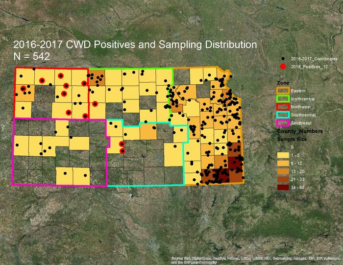
0 Comments:
Post a Comment
Subscribe to Post Comments [Atom]
<< Home