TEXAS CWD TSE PRION REPORTING TURKEY OF THE YEAR AWARD GOES TO SHANNON
TOMPKINS OF THE HOUSTON CHRONICLE
Once again, the legendary and award winning outdoors writer Shannon
Tompkins of the Houston Chronicle writes another great article of how blessed we
are to be in Texas ;
Deer season giving hunters plenty of reasons to be thankful
By Shannon Tompkins Updated 8:45 pm,
Wednesday, November 25, 2015
‘’and those deer hunters have plenty for which to be thankfully
optimistic’’.
Then the rest of Mr. Tompkins full page article (except for add) goes on to
tell a fascinating tale of the bountiful fruits of the hunt across Texas, and
what this years fruits of the hunt might hold. It was another great article.
But, once again, for reasons I can only imagine (industry), what Shannon Tomkins
and the Houston Chronicle failed to do, they failed to remind it’s readers that
everything across the great state of Texas is not as rosy as they proclaim it to
be this hunting season. CHRONIC WASTING DISEASE CWD TSE PRION aka mad deer
disease, has now been documented in Texas in the wild and captive cervid. Cases
are mounting in both wild and captive deer, scientist are showing much concern
about the potential from exposure to the CWD TSE Prion in humans and other
species (PRION2015 conference), and the threat to Texas not only to humans and
other species, but to the environment is very real, and could have horrible and
detrimental long lasting effects. It would seem to me, that since TEXAS PARKS
AND WILDLIFE DEPARTMENT EXECUTIVE DIRECTOR ORDER NO. 015-006 Chronic Wasting
Disease (CWD) IMMEDIATE DANGER TO THE WHILE-TAILED DEER AND MULE DEER RESOURCES
OF TEXAS was just declared October 5, 2015, and the dire straights this bings,
NONE of this was mentioned is this article. In my opinion, it’s not only
appalling, but it’s beginning to paint a broader picture in my mind, but I will
leave that picture in my mind for now. While the state of Michigan is waging a
similar battle with CWD TSE Prion, in Michigan, they go to great efforts to try
and bring awareness and these risk factors to the public, with a program KEEP
THE U.P. CWD FREE!!
Michigan hands out bumper stickers and pass out written brochure to the
public trying to bring awareness to the CWD TSE Prion.
while in Texas, it’s just the opposite, it’s all hush hush, we are still
waiting with great anticipation on the latest two suspect CWD cases breeder
captive, the silence is still deafening ???
By Pilar Arias - Reporter , David Ibanez - Web - Managing Editor
Posted: 10:12 AM, November 14, 2015
Updated: 10:13 AM, November 14, 2015
Two new cases of Chronic Wasting Disease CWD in captive white-tailed deer
have been reported, a Texas Parks and Wildlife Department official said. There
have now been seven cases of the fatal disease reported in Texas...
Saturday, November 14, 2015
TEXAS CAPTIVE BREEDER CHRONIC WASTING DISEASE CWD 2 MORE SUSPECTS DECTECTED
where are any statements from the TAHC or TPWD either confirming this, or
refuting this???
actually, if these two new captive suspect CWD cases are confirmed, that
would be a total of 7 cases of CWD in Captive in Texas, PLUS the 8 other
confirmed cases of CWD up in the Texas Trans Pecos region to date in the mule
deer. So the total would be 15 cases of the CWD TSE Prion aka mad cow type
disease in Cervid in Texas, to date. just to put everything in perspective. BUT,
that would only be IF and WHEN, the TAHC or the TPWD ever confirm these two new
recent suspect CWD cases.
I am only reminded of another great article Shannon Tompkins wrote years
ago, when the CWD TSE Prion shoe was on the other foot...
March 14, 2002
"Ten years ago, elk and deer (imported into Texas) were not regulated at
all," said Dr. Ken Waldrup, an epidemiologist with the Texas Animal Health
Commission and one of the agency's point men on CWD. "If Texas doesn't already
have CWD, then I say that proves that God is a Texan. "For everyone's sake, I
sure hope He is."
========================
*** Tompkins: There are a lot of reasons to be concerned about CWD
Houston Chronicle Published 5:30 a.m., Thursday, March 14, 2002
Today, most Texas deer hunters probably yawn at the mention of Chronic
Wasting Disease. After all, the number of wild deer documented as killed,
nationwide, by the unusual malady probably is less than annually are crushed by
tractor-trailer rigs scorching Interstate 10 between Kerrville and Fort
Stockton.
And, so far, no cases of the fatal, incurable, communicable,
brain-destroying cervid disease have been documented in Texas.
What's so bad about a little-understood disease responsible for the death
of scattered pockets of deer in a handful of Rocky Mountain states? If Texas'
deer herd survived screwworms and can thrive despite endemic bluetongue and
anthrax and even the constant gnawing away of habitat, then why worry about a
little Chronic Wasting Disease?
*** There is abundant reason to be concerned.
*** CWD carries potential for incredible impacts on Texas' 4 million deer,
its half-million deer hunters, the hunting-based economy of rural areas and
private landowners and even the future of the state agency responsible for
overseeing those deer and all other natural resources.
Just how seriously many Texas wildlife managers and those with economic or
other interest in deer take the CWD threat was manifestly evident over the past
week.
In the wake of news that Wisconsin officials had discovered CWD in three of
26 wild deer taken by hunters in a small area of that state, Katharine Armstrong
Idsal, presiding officer of the Texas Parks and Wildlife Commission, called an
emergency meeting of the TPW Commission to address the issue of deer importation
into Texas.
A proposal to suspend all imports of deer into Texas was, and is, on the
TPW Commission's agenda for its scheduled April 4 meeting, with the
recommendation having been triggered by discovery over the past few months of
CWD in wild deer in Nebraska and South Dakota.
The emergency TPW Commission meeting was arranged Friday, the day the Texas
Wildlife Association, a politically active, landowner-based organization, sent
to the governor, members of the Legislature and the TPW Commission a resolution
calling for sealing the state's borders to deer imports because of the chance
some might be carrying CWD.
At the TPW Commission's hastily called Monday meeting, the group approved
and adopted an emergency rule prohibiting importation of white-tailed and mule
deer into Texas.
That emergency rule, which is effective for 45 days, took effect Tuesday.
It is the first time the TPW Commission has used its emergency rule-making
authority.
Justifications for the emergency action were laid out in the preamble to
the regulation change. CWD, the document states, "constitutes a direct threat to
wild deer populations in Texas and therefore to the multi-billion dollar hunting
industry, as well as a potential threat to human health, safety and welfare."
To understand the threat to deer and, perhaps, public health and the
subsequent potentially devastating impact on Texas' deer-based economy, it's
necessary to understand CWD.
CWD is one of a group of transmissible spongiform encephalopathies (TSE)
diseases that destroy brain cells. Triggering the destruction is a prion, an
abnormal form of protein. The prion mutates normal cellular protein into the
abnormal form.
This "eats away" at the brain and damages an infected animal's ability to
maintain normal functions such as converting food and body fat to energy.
Animals suffering from CWD begin wasting away as their body tries to
convert protein to energy, a very inefficient process.
Eventually, the animal loses motor control and even goes blind, giving rise
to the pitiful "blind staggers" seen in livestock suffering from CWD's close
relative, Bovine Spongiform Encephalopathy, better known as "Mad Cow Disease."
Death is inevitable and horrible.
Scientists know relatively little about CWD.
"We don't really know what triggers it. Does the prion create the disease
or does the disease create the prion?" said Jerry Cooke, game mammal branch
chief of the Texas Parks and Wildlife Department's wildlife division. "What we
do know is that it is transmissible to other cervids."
First documented in the 1960s in penned herds in Colorado, CWD "jumped"
into the wild cervid population there, being confirmed in wild deer and elk in
the 1980s.
A common suspicion is that CWD is a mutated form of "scrapie," a TSE long
confined to sheep.
There is some evidence that the cervids in the Colorado pen where CWD was
first documented were fed protein feeds containing sheep parts and that those
parts could have contained brain material infected with scrapie.
One of the scrapie-triggering prions might have mutated just enough to
break the molecular barrier of a deer's brain cell, and the disease was off and
running.
Scientists are convinced CWD is spread by close contact between uninfected
and infected animals. That can happen between animals in a pen or behind a
fence, or by nose-to-nose contact between deer or elk inside the fence and those
outside the enclosure.
From Colorado, CWD spread throughout the northwest corner of the state into
wild herds in Wyoming and Nebraska.
Its spread was accelerated over the past decade by a burgeoning market in
deer and elk triggered by elk farming and deer ranching.
Thousands of deer and elk are bought and transported each year, most to
penned facilities where they are either raised for food or, in the case of
white-tailed deer, used in an effort to produce bucks with large antlers to feed
a market in trophy hunting.
To test for CWD, brain tissue is needed. And such tissue samples can be
obtained only if the animal is dead.
Plus, getting rid of the disease has proved difficult, if not impossible,
even in penned facilities.
In at least one case, a penned facility holding CWD-infected deer was
"depopulated" (the animals slaughtered and destroyed) and the site left with no
animals for three years.
When uninfected deer were placed in the pens, they contracted CWD.
As deer and elk from areas with CWD have been traded and transported across
the nation, they have brought the disease with them
Currently, CWD-infected, free-ranging deer have been confirmed in Colorado,
Nebraska, Wyoming, South Dakota and Wisconsin, plus the Canadian province of
Saskatchewan.
CWD has been found in captive herds in Saskatchewan, Colorado, South
Dakota, Nebraska, Kansas, Oklahoma and Montana.
Texas has been a big player in the deer trade over the past decade, as
hundreds of deer-breeding facilities have sprung up in the state to feed the
interest in building bucks with bigger antlers.
Today, more than 450 individuals in Texas hold a TPWD-issued "scientific
breeder permit" allowing them to manipulate deer. Some of these breeders and
other landowners over the past four years have imported 2,107 deer from outside
Texas.
Because deer can be traded so often -- a deer may be sold as a fawn in
Nebraska to a broker in Missouri who sells it to a breeder in Pennsylvania who
sells it to a landowner in Texas -- it often is nearly impossible to determine
the provenance of individual animals.
Whether any of the thousands of deer imported into Texas over the past
decade carried CWD remains an unsettling question.
Texas has no CWD-testing program for wild deer and only a voluntary program
for elk and other animals under the jurisdiction of the Texas Animal Health
Commission.
"Ten years ago, elk and deer (imported into Texas) were not regulated at
all," said Dr. Ken Waldrup, an epidemiologist with the Texas Animal Health
Commission and one of the agency's point men on CWD. "If Texas doesn't already
have CWD, then I say that proves that God is a Texan.
"For everyone's sake, I sure hope He is."
CWD has not been proved to be transmissible to any animal other than deer
and elk.
But that was the original thought with BSE, which did "jump" into humans
who ate BSE-infected meat in Europe and contracted Creutzfeldt-Jakob Disease
(CJD), the human form of TSE. CJD, like CWD and BSE, is fatal, incurable and
untreatable. It is blamed for at least 80 deaths in Europe.
While there is no proof CWD can jump to humans, there is no absolute proof
it can't if given enough opportunities.
And that issue scares wildlife managers.
If CWD shows up in a deer herd and the deer-hunting public gets spooked
about the possibility -- no matter how tiny -- that by cleaning or eating a deer
they will contract CJD and face a certain and horrible death, they could, en
masse, abandon deer hunting.
This could destroy the $2 billion-plus deer hunting economy in Texas.
Also, if deer hunters abandon their recreation, natural resource agencies
such as TPWD, which depend almost entirely on hunting license fees to fund their
diverse wildlife programs, would be maimed, perhaps mortally.
"It's not the immediate impact on the deer herds that (is) the most
frightening thing about CWD," Waldrup said. "It's the secondary impacts that are
really scary.
"People better just pray it doesn't show up here. If it does, things could
get very ugly."
Shannon Tompkins covers the outdoors for the Chronicle. His column appears
Thursdays, Fridays and Sundays.
Saturday, October 03, 2015
TEXAS CHRONIC WASTING DISEASE CWD TSE PRION GOD MUST NOT BE A TEXAN 2002 TO
2015
Monday, November 16, 2015
*** TEXAS PARKS AND WILDLIFE DEPARTMENT EXECUTIVE DIRECTOR ORDER NO.
015-006
*** Chronic Wasting Disease (CWD) immediate danger to the white-tailed deer
and mule deer resources of Texas
Thursday, November 05, 2015
*** TPW Commission Adopts Interim Deer Breeder Movement Rules
Friday, October 09, 2015
Texas TWA Chronic Wasting Disease TSE Prion Webinars and Meeting October
2015
Thursday, September 24, 2015
TEXAS Hunters Asked to Submit Samples for Chronic Wasting Disease CWD TSE
Prion Testing
*** I cannot stress enough to all of you, for the sake of your family and
mine, before putting anything in the freezer, have those deer tested for CWD.
...terry
Friday, August 07, 2015
*** Texas CWD Captive, and then there were 4 ?
Thursday, August 06, 2015
*** WE HAVE LOST TEXAS TO CWD TASK FORCE CATERING TO INDUSTRY
Tuesday, July 21, 2015
*** Texas CWD Medina County Herd Investigation Update July 16, 2015 ***
Wednesday, July 01, 2015
*** TEXAS Chronic Wasting Disease Detected in Medina County Captive Deer
Wednesday, March 25, 2015
*** Chronic Wasting Disease CWD Cases Confirmed In New Mexico 2013 and 2014
UPDATE 2015
Wednesday, March 18, 2015
*** Chronic Wasting Disease CWD Confirmed Texas Trans Pecos March 18, 2015
Monday, February 11, 2013
TEXAS CHRONIC WASTING DISEASE CWD Four New Positives Found in Trans Pecos
Tuesday, July 10, 2012
Chronic Wasting Disease Detected in Far West Texas
July 10, 2012
Chronic Wasting Disease Detected in Far West Texas
AUSTIN -- Samples from two mule deer recently taken in far West Texas have
been confirmed positive for Chronic Wasting Disease (CWD). These are the first
cases of CWD detected in Texas deer. Wildlife officials believe the event is
currently isolated in a remote part of the state near the New Mexico border.
CENSORED, RAW, AND UNCUT...
Sunday, August 23, 2015
*** TAHC Chronic Wasting Disease CWD TSE Prion and how to put lipstick on a
pig and take her to the dance in Texas
from the other side of the fence... today’s Singeltary Sunday School class
‘thinking outside of the box, God’s Wrath’ at the bottom. ...tss
Thursday, March 14, 2013
*** TEXAS DEER BREEDERS CHEER TWO NEW BILLS SB 1444 AND HB 2092 THAT COULD
HELP POTENTIALLY ENHANCE CHRONIC WASTING DISEASE CWD ***
Friday, August 14, 2015
*** Susceptibility of cattle to the agent of chronic wasting disease from
elk after intracranial inoculation
Friday, August 14, 2015
*** Carcass Management During a Mass Animal Health Emergency Draft
Programmatic Environmental Impact Statement—August 2015
Tuesday, September 22, 2015
*** Host Determinants of Prion Strain Diversity Independent of Prion
Protein Genotype
Friday, August 28, 2015
*** Chronic Wasting Disease CWD TSE Prion Diagnostics and subclinical
infection ***
Research Project: TRANSMISSION, DIFFERENTIATION, AND PATHOBIOLOGY OF
TRANSMISSIBLE SPONGIFORM ENCEPHALOPATHIES
*** Title: Transmission of scrapie prions to primate after an extended
silent incubation period Authors
item Comoy, Emmanuel - item Mikol, Jacqueline - item Luccantoni-Freire,
Sophie - item Correia, Evelyne - item Lescoutra-Etchegaray, Nathalie - item
Durand, Valérie - item Dehen, Capucine - item Andreoletti, Olivier - item
Casalone, Cristina - item Richt, Juergen item Greenlee, Justin item Baron,
Thierry - item Benestad, Sylvie - item Hills, Bob - item Brown, Paul - item
Deslys, Jean-Philippe -
Submitted to: Scientific Reports Publication Type: Peer Reviewed Journal
Publication Acceptance Date: May 28, 2015 Publication Date: June 30, 2015
Citation: Comoy, E.E., Mikol, J., Luccantoni-Freire, S., Correia, E.,
Lescoutra-Etchegaray, N., Durand, V., Dehen, C., Andreoletti, O., Casalone, C.,
Richt, J.A., Greenlee, J.J., Baron, T., Benestad, S., Brown, P., Deslys, J.
2015. Transmission of scrapie prions to primate after an extended silent
incubation period. Scientific Reports. 5:11573. Interpretive Summary: The
transmissible spongiform encephalopathies (also called prion diseases) are fatal
neurodegenerative diseases that affect animals and humans. The agent of prion
diseases is a misfolded form of the prion protein that is resistant to breakdown
by the host cells. Since all mammals express prion protein on the surface of
various cells such as neurons, all mammals are, in theory, capable of
replicating prion diseases. One example of a prion disease, bovine spongiform
encephalopathy (BSE; also called mad cow disease), has been shown to infect
cattle, sheep, exotic undulates, cats, non-human primates, and humans when the
new host is exposed to feeds or foods contaminated with the disease agent. The
purpose of this study was to test whether non-human primates (cynomologous
macaque) are susceptible to the agent of sheep scrapie. After an incubation
period of approximately 10 years a macaque developed progressive clinical signs
suggestive of neurologic disease. Upon postmortem examination and microscopic
examination of tissues, there was a widespread distribution of lesions
consistent with a transmissible spongiform encephalopathy. This information will
have a scientific impact since it is the first study that demonstrates the
transmission of scrapie to a non-human primate with a close genetic relationship
to humans. This information is especially useful to regulatory officials and
those involved with risk assessment of the potential transmission of animal
prion diseases to humans.
Technical Abstract: Classical bovine spongiform encephalopathy (c-BSE) is
an animal prion disease that also causes variant Creutzfeldt-Jakob disease in
humans. Over the past decades, c-BSE's zoonotic potential has been the driving
force in establishing extensive protective measures for animal and human health.
In complement to the recent demonstration that humanized mice are susceptible to
scrapie, we report here the first observation of direct transmission of a
natural classical scrapie isolate to a macaque after a 10-year incubation
period. Neuropathologic examination revealed all of the features of a prion
disease: spongiform change, neuronal loss, and accumulation of PrPres throughout
the CNS.
***This observation strengthens the questioning of the harmlessness of
scrapie to humans, at a time when protective measures for human and animal
health are being dismantled and reduced as c-BSE is considered controlled and
being eradicated. Our results underscore the importance of precautionary and
protective measures and the necessity for long-term experimental transmission
studies to assess the zoonotic potential of other animal prion strains.
Research Project: TRANSMISSION, DIFFERENTIATION, AND PATHOBIOLOGY OF
TRANSMISSIBLE SPONGIFORM ENCEPHALOPATHIES
Title: Transmission of the agent of sheep scrapie to deer results in PrPSc
with two distinct molecular profiles Authors
item Greenlee, Justin item Moore, Sarah - item Smith, Jodi item West
Greenlee, Mary - item Kunkle, Robert
Submitted to: Prion Publication Type: Abstract Only Publication Acceptance
Date: March 31, 2015 Publication Date: May 25, 2015 Citation: Greenlee, J.,
Moore, S.J., Smith, J.., West Greenlee, M.H., Kunkle, R. 2015.
Scrapie transmits to white-tailed deer by the oral route and has a
molecular profile similar to chronic wasting disease and distinct from the
scrapie inoculum. Prion 2015. p. S62. Technical Abstract: The purpose of this
work was to determine susceptibility of white-tailed deer (WTD) to the agent of
sheep scrapie and to compare the resultant PrPSc to that of the original
inoculum and chronic wasting disease (CWD). We inoculated WTD by a natural route
of exposure (concurrent oral and intranasal (IN); n=5) with a US scrapie
isolate. All scrapie-inoculated deer had evidence of PrPSc accumulation. PrPSc
was detected in lymphoid tissues at preclinical time points, and deer necropsied
after 28 months post-inoculation had clinical signs, spongiform encephalopathy,
and widespread distribution of PrPSc in neural and lymphoid tissues. Western
blotting (WB) revealed PrPSc with 2 distinct molecular profiles. WB on cerebral
cortex had a profile similar to the original scrapie inoculum, whereas WB of
brainstem, cerebellum, or lymph nodes reveal PrPSc with a higher profile
resembling CWD. Homogenates with the 2 distinct profiles from WTD with clinical
scrapie were further passaged to mice expressing cervid prion protein and
intranasally to sheep and WTD. In cervidized mice, the two inocula have distinct
incubation times. Sheep inoculated intranasally with WTD derived scrapie
developed disease, but only after inoculation with the inoculum that had a
scrapie-like profile. The WTD study is ongoing, but deer in both inoculation
groups are positive for PrPSc by rectal mucosal biopsy. In summary, this work
demonstrates that WTD are susceptible to the agent of scrapie, two distinct
molecular profiles of PrPSc are present in the tissues of affected deer, and
inoculum of either profile type readily passes to deer.
Docket No. APHIS-2007-0127 Scrapie in Sheep and Goats Terry Singeltary Sr.
Submission
Docket No. APHIS-2007-0127 Scrapie in Sheep and Goats
SUMMARY: We are reopening the comment period for our proposed rule that
would revise completely the scrapie regulations, which concern the risk groups
and categories established for individual animals and for flocks, the use of
genetic testing as a means of assigning risk levels to animals, movement
restrictions for animals found to be genetically less susceptible or resistant
to scrapie, and recordkeeping requirements. This action will allow interested
persons additional time to prepare and submit comments.DATES: The comment period
for the proposed rule published on September 10, 2015 (80 FR 54660-54692) is
reopened. We will consider all comments that we receive on or before December 9,
2015. ...
COMMENT SUBMISSION TERRY S. SINGELTARY SR.
WITH regards to Docket No. APHIS-2007-0127 Scrapie in Sheep and Goats, I
kindly submit the following ;
>>>The last major revision of the scrapie regulations occurred on
August 21, 2001, when we published in theFederal Register(66 FR 43964, Docket
No. 97-093-5) a final rule amending part 79 by imposing additional restrictions
on the interstate movement of sheep and goats.<<<
Indeed, much science has changed about the Scrapie TSE prion, including
more science linking Scrapie to humans. sadly, politics, industry, and trade,
have not changed, and those usually trump sound science, as is the case with all
Transmissible Spongiform Encephalopathy TSE Prion disease in livestock producing
animals and the OIE. we can look no further at the legal trading of the Scrapie
TSE prion both typical and atypical of all strains, and CWD all stains. With as
much science of old, and now more new science to back this up, Scrapie of all
types i.e. atypical and typical, BSE all strains, and CWD all strains, should be
regulated in trade as BSE TSE PRION. In fact, I urge APHIS et al and the OIE,
and all trading partners to take heed to the latest science on the TSE prion
disease, all of them, and seriously reconsider the blatant disregards for human
and animal health, all in the name of trade, with the continued relaxing of TSE
Prion trade regulations through the ‘NEGLIGIBLE BSE RISK’ PROGRAM, which was set
up to fail in the first place. If the world does not go back to the ‘BSE RISK
ASSESSMENTS’, enhance, and or change that assessment process to include all TSE
prion disease, i.e. ‘TSE RISK ASSESSMENT’, if we do not do this and if we
continue this farce with OIE and the USDA et al, and the ‘NEGLIGIBLE BSE RISK’
PROGRAM, we will never eradicate the TSE prion aka mad cow type disease, they
will continue to mutate and spread among species of human and animal origin, and
they will continue to kill. ...
please see ;
O.05: Transmission of prions to primates after extended silent incubation
periods: Implications for BSE and scrapie risk assessment in human populations
Emmanuel Comoy, Jacqueline Mikol, Val erie Durand, Sophie Luccantoni,
Evelyne Correia, Nathalie Lescoutra, Capucine Dehen, and Jean-Philippe Deslys
Atomic Energy Commission; Fontenay-aux-Roses, France
Prion diseases (PD) are the unique neurodegenerative proteinopathies
reputed to be transmissible under field conditions since decades. The
transmission of Bovine Spongiform Encephalopathy (BSE) to humans evidenced that
an animal PD might be zoonotic under appropriate conditions. Contrarily, in the
absence of obvious (epidemiological or experimental) elements supporting a
transmission or genetic predispositions, PD, like the other proteinopathies, are
reputed to occur spontaneously (atpical animal prion strains, sporadic CJD
summing 80% of human prion cases). Non-human primate models provided the first
evidences supporting the transmissibiity of human prion strains and the zoonotic
potential of BSE. Among them, cynomolgus macaques brought major information for
BSE risk assessment for human health (Chen, 2014), according to their
phylogenetic proximity to humans and extended lifetime. We used this model to
assess the zoonotic potential of other animal PD from bovine, ovine and cervid
origins even after very long silent incubation periods.
*** We recently observed the direct transmission of a natural classical
scrapie isolate to macaque after a 10-year silent incubation period,
***with features similar to some reported for human cases of sporadic CJD,
albeit requiring fourfold longe incubation than BSE. Scrapie, as recently evoked
in humanized mice (Cassard, 2014),
***is the third potentially zoonotic PD (with BSE and L-type BSE),
***thus questioning the origin of human sporadic cases. We will present an
updated panorama of our different transmission studies and discuss the
implications of such extended incubation periods on risk assessment of animal PD
for human health.
===============
***thus questioning the origin of human sporadic cases***
===============
***This information will have a scientific impact since it is the first
study that demonstrates the transmission of scrapie to a non-human primate with
a close genetic relationship to humans. This information is especially useful to
regulatory officials and those involved with risk assessment of the potential
transmission of animal prion diseases to humans.
***This observation strengthens the questioning of the harmlessness of
scrapie to humans, at a time when protective measures for human and animal
health are being dismantled and reduced as c-BSE is considered controlled and
being eradicated. Our results underscore the importance of precautionary and
protective measures and the necessity for long-term experimental transmission
studies to assess the zoonotic potential of other animal prion strains.
Monday, November 16, 2015
*** Docket No. APHIS-2007-0127 Scrapie in Sheep and Goats Terry Singeltary
Sr. Submission ***
Tuesday, September 29, 2015
*** Transmission of chronic wasting disease to sentinel reindeer (Rangifer
tarandus tarandus) can transmit CWD to naive reindeer both directly and
indirectly ***
Research Project: TRANSMISSION, DIFFERENTIATION, AND PATHOBIOLOGY OF
TRANSMISSIBLE SPONGIFORM ENCEPHALOPATHIES
Transmission of chronic wasting disease to sentinel reindeer (Rangifer
tarandus tarandus) can transmit CWD to naive reindeer both directly and
indirectly Research Project: TRANSMISSION, DIFFERENTIATION, AND PATHOBIOLOGY OF
TRANSMISSIBLE SPONGIFORM ENCEPHALOPATHIES
Title: Transmission of chronic wasting disease to sentinel reindeer
(Rangifer tarandus tarandus)
Authors item Moore, S - item Kunkle, Robert item Nicholson, Eric item
Richt, Juergen item Hamir, Amirali item Waters, Wade item Greenlee, Justin
Submitted to: American College of Veterinary Pathologists Meeting Publication
Type: Abstract Only Publication Acceptance Date: August 12, 2015 Publication
Date: N/A Technical
Abstract: Chronic wasting disease (CWD) is a naturally-occurring, fatal
neurodegenerative disease of North American cervids. Reindeer (Rangifer tarandus
tarandus) are susceptible to CWD following oral challenge, but CWD has not been
reported in free-ranging caribou (Rangifer tarandus caribou) or farmed reindeer.
Potential contact between CWD-affected cervids and Rangifer species that are
free-ranging or co-housed on farms presents a potential risk of CWD
transmission. The aims of this study were to 1) investigate the transmission of
CWD from white-tailed deer (Odocoileus virginianus; CWD-wtd), mule deer
(Odocoileus hemionus; CWD-md), or elk (Cervus elaphus nelsoni; CWD-elk) to
reindeer via the intracranial route, and 2) to assess for direct and indirect
horizontal transmission to non-inoculated sentinels. Three groups of 5 reindeer
fawns were challenged intracranially with CWD-wtd, CWD-md, or CWD-elk. Two years
after challenge of inoculated reindeer, non-inoculated control reindeer were
introduced into the same pen as the CWD-wtd inoculated reindeer (n=4) or into a
pen adjacent to the CWD-md inoculated reindeer (n=2). Reindeer were allowed to
develop clinical disease. At death/euthanasia a complete necropsy examination
was performed, including immunohistochemical testing of tissues for
disease-associated CWD prion protein (PrP-CWD). Intracranially challenged
reindeer developed clinical disease from 21 months post-inoculation (MPI).
***PrP-CWD was detected in 5/6 sentinel reindeer although only 2/6
developed clinical disease during the study period (<57 and="" are="" both="" can="" cervid="" cwd="" directly="" div="" from="" have="" indirectly.="" mpi="" naive="" reindeer="" shown="" sources="" susceptible="" that="" to="" transmit="" various="" we="">
the tse prion aka mad cow type disease is not your normal pathogen.
The TSE prion disease survives ashing to 600 degrees celsius, that’s around
1112 degrees farenheit.
you cannot cook the TSE prion disease out of meat. you can take the ash and
mix it with saline and inject that ash into a mouse, and the mouse will go down
with TSE.
Prion Infected Meat-and-Bone Meal Is Still Infectious after Biodiesel
Production as well.
the TSE prion agent also survives Simulated Wastewater Treatment Processes.
IN fact, you should also know that the TSE Prion agent will survive in the
environment for years, if not decades.
you can bury it and it will not go away.
The TSE agent is capable of infected your water table i.e. Detection of
protease-resistant cervid prion protein in water from a CWD-endemic area.
it’s not your ordinary pathogen you can just cook it out and be done with.
that’s what’s so worrisome about Iatrogenic mode of transmission, a simple
autoclave will not kill this TSE prion agent.
cwd to humans, consumption, exposure, sub-clinical, iatrogenic, what if ?
Tuesday, September 29, 2015
*** Transmission of chronic wasting disease to sentinel reindeer (Rangifer
tarandus tarandus) can transmit CWD to naive reindeer both directly and
indirectly
Research Project: TRANSMISSION, DIFFERENTIATION, AND PATHOBIOLOGY OF
TRANSMISSIBLE SPONGIFORM ENCEPHALOPATHIES
*** Infectious agent of sheep scrapie may persist in the environment for at
least 16 years ***
Gudmundur Georgsson1, Sigurdur Sigurdarson2 and Paul Brown3
*** Spraker suggested an interesting explanation for the occurrence of CWD.
The deer pens at the Foot Hills Campus were built some 30-40 years ago by a Dr.
Bob Davis. At or abut that time, allegedly, some scrapie work was conducted at
this site. When deer were introduced to the pens they occupied ground that had
previously been occupied by sheep.
HIGHEST INFECTION RATE ON SEVERAL CWD CONFIRMED CAPTIVES
CHRONIC WASTING DISEASE CWD WISCONSIN Almond Deer (Buckhorn Flats) Farm
Update DECEMBER 2011
The CWD infection rate was nearly 80%, the highest ever in a North American
captive herd.
RECOMMENDATION: That the Board approve the purchase of 80 acres of land for
$465,000 for the Statewide Wildlife Habitat Program in Portage County and
approve the restrictions on public use of the site.
SUMMARY:
For Immediate Release Thursday, October 2, 2014
Dustin Vande Hoef 515/281-3375 or 515/326-1616 (cell) or
Dustin.VandeHoef@IowaAgriculture.gov
*** TEST RESULTS FROM CAPTIVE DEER HERD WITH CHRONIC WASTING DISEASE
RELEASED 79.8 percent of the deer tested positive for the disease
DES MOINES – The Iowa Department of Agriculture and Land Stewardship today
announced that the test results from the depopulation of a quarantined captive
deer herd in north-central Iowa showed that 284 of the 356 deer, or 79.8% of the
herd, tested positive for Chronic Wasting Disease (CWD).
*** see history of this CWD blunder here ;
On June 5, 2013, DNR conducted a fence inspection, after gaining approval
from surrounding landowners, and confirmed that the fenced had been cut or
removed in at least four separate locations; that the fence had degraded and was
failing to maintain the enclosure around the Quarantined Premises in at least
one area; that at least three gates had been opened;and that deer tracks were
visible in and around one of the open areas in the sand on both sides of the
fence, evidencing movement of deer into the Quarantined Premises.
The overall incidence of clinical CWD in white-tailed deer was 82%
Species (cohort) CWD (cases/total) Incidence (%) Age at CWD death (mo)
Thursday, November 19, 2015
Wisconsin Eau Claire Co. deer herd two day round of depopulation CWD
testing shows 23 positive
”The occurrence of CWD must be viewed against the contest of the locations
in which it occurred. It was an incidental and unwelcome complication of the
respective wildlife research programmes. Despite it’s subsequent recognition as
a new disease of cervids, therefore justifying direct investigation, no specific
research funding was forthcoming. The USDA veiwed it as a wildlife problem and
consequently not their province!” page 26.
Sunday, January 06, 2013
USDA TO PGC ONCE CAPTIVES ESCAPE
*** "it‘s no longer its business.”
CWD, spreading it around...
for the game farm industry, and their constituents, to continue to believe
that they are _NOT_, and or insinuate that they have _NEVER_ been part of the
problem, will only continue to help spread cwd. the game farming industry, from
the shooting pens, to the urine mills, the antler mills, the sperm mills, velvet
mills, shooting pens, to large ranches, are not the only problem, but it is
painfully obvious that they have been part of the problem for decades and
decades, just spreading it around, as with transportation and or exportation and
or importation of cervids from game farming industry, and have been proven to
spread cwd. no one need to look any further than South Korea blunder ;
===========================================
spreading cwd around...
Between 1996 and 2002, chronic wasting disease was diagnosed in 39 herds of
farmed elk in Saskatchewan in a single epidemic. All of these herds were
depopulated as part of the Canadian Food Inspection Agency’s (CFIA) disease
eradication program. Animals, primarily over 12 mo of age, were tested for the
presence CWD prions following euthanasia. Twenty-one of the herds were linked
through movements of live animals with latent CWD from a single infected source
herd in Saskatchewan, 17 through movements of animals from 7 of the secondarily
infected herds.
***The source herd is believed to have become infected via importation of
animals from a game farm in South Dakota where CWD was subsequently diagnosed
(7,4). A wide range in herd prevalence of CWD at the time of herd depopulation
of these herds was observed. Within-herd transmission was observed on some
farms, while the disease remained confined to the introduced animals on other
farms.
spreading cwd around...
Friday, May 13, 2011
Chronic Wasting Disease (CWD) outbreaks and surveillance program in the
Republic of Korea
Hyun-Joo Sohn, Yoon-Hee Lee, Min-jeong Kim, Eun-Im Yun, Hyo-Jin Kim,
Won-Yong Lee, Dong-Seob Tark, In- Soo Cho, Foreign Animal Disease Research
Division, National Veterinary Research and Quarantine Service, Republic of Korea
Chronic wasting disease (CWD) has been recognized as an important prion
disease in native North America deer and Rocky mountain elks. The disease is a
unique member of the transmissible spongiform encephalopathies (TSEs), which
naturally affects only a few species. CWD had been limited to USA and Canada
until 2000.
On 28 December 2000, information from the Canadian government showed that a
total of 95 elk had been exported from farms with CWD to Korea. These consisted
of 23 elk in 1994 originating from the so-called “source farm” in Canada, and 72
elk in 1997, which had been held in pre export quarantine at the “source
farm”.Based on export information of CWD suspected elk from Canada to Korea, CWD
surveillance program was initiated by the Ministry of Agriculture and Forestry
(MAF) in 2001.
All elks imported in 1997 were traced back, however elks imported in 1994
were impossible to identify. CWD control measures included stamping out of all
animals in the affected farm, and thorough cleaning and disinfection of the
premises. In addition, nationwide clinical surveillance of Korean native
cervids, and improved measures to ensure reporting of CWD suspect cases were
implemented.
Total of 9 elks were found to be affected. CWD was designated as a
notifiable disease under the Act for Prevention of Livestock Epidemics in 2002.
Additional CWD cases - 12 elks and 2 elks - were diagnosed in 2004 and
2005.
Since February of 2005, when slaughtered elks were found to be positive,
all slaughtered cervid for human consumption at abattoirs were designated as
target of the CWD surveillance program. Currently, CWD laboratory testing is
only conducted by National Reference Laboratory on CWD, which is the Foreign
Animal Disease Division (FADD) of National Veterinary Research and Quarantine
Service (NVRQS).
In July 2010, one out of 3 elks from Farm 1 which were slaughtered for the
human consumption was confirmed as positive. Consequently, all cervid – 54 elks,
41 Sika deer and 5 Albino deer – were culled and one elk was found to be
positive. Epidemiological investigations were conducted by Veterinary
Epidemiology Division (VED) of NVRQS in collaboration with provincial veterinary
services.
Epidemiologically related farms were found as 3 farms and all cervid at
these farms were culled and subjected to CWD diagnosis. Three elks and 5
crossbreeds (Red deer and Sika deer) were confirmed as positive at farm 2.
All cervids at Farm 3 and Farm 4 – 15 elks and 47 elks – were culled and
confirmed as negative.
Further epidemiological investigations showed that these CWD outbreaks were
linked to the importation of elks from Canada in 1994 based on circumstantial
evidences.
In December 2010, one elk was confirmed as positive at Farm 5.
Consequently, all cervid – 3 elks, 11 Manchurian Sika deer and 20 Sika deer –
were culled and one Manchurian Sika deer and seven Sika deer were found to be
positive. This is the first report of CWD in these sub-species of deer.
Epidemiological investigations found that the owner of the Farm 2 in CWD
outbreaks in July 2010 had co-owned the Farm 5.
In addition, it was newly revealed that one positive elk was introduced
from Farm 6 of Jinju-si Gyeongsang Namdo. All cervid – 19 elks, 15 crossbreed
(species unknown) and 64 Sika deer – of Farm 6 were culled, but all confirmed as
negative.
New studies on the heat resistance of hamster-adapted scrapie agent:
Threshold survival after ashing at 600°C suggests an inorganic template of
replication
The infectious agents responsible for transmissible spongiform
encephalopathy (TSE) are notoriously resistant to most physical and chemical
methods used for inactivating pathogens, including heat. It has long been
recognized, for example, that boiling is ineffective and that higher
temperatures are most efficient when combined with steam under pressure (i.e.,
autoclaving). As a means of decontamination, dry heat is used only at the
extremely high temperatures achieved during incineration, usually in excess of
600°C. It has been assumed, without proof, that incineration totally inactivates
the agents of TSE, whether of human or animal origin.
Prion Infected Meat-and-Bone Meal Is Still Infectious after Biodiesel
Production
Histochemical analysis of hamster brains inoculated with the solid residue
showed typical spongiform degeneration and vacuolation. Re-inoculation of these
brains into a new cohort of hamsters led to onset of clinical scrapie symptoms
within 75 days, suggesting that the specific infectivity of the prion protein
was not changed during the biodiesel process. The biodiesel reaction cannot be
considered a viable prion decontamination method for MBM, although we observed
increased survival time of hamsters and reduced infectivity greater than 6 log
orders in the solid MBM residue. Furthermore, results from our study compare for
the first time prion detection by Western Blot versus an infectivity bioassay
for analysis of biodiesel reaction products. We could show that biochemical
analysis alone is insufficient for detection of prion infectivity after a
biodiesel process.
Detection of protease-resistant cervid prion protein in water from a
CWD-endemic area
The data presented here demonstrate that sPMCA can detect low levels of
PrPCWD in the environment, corroborate previous biological and experimental data
suggesting long term persistence of prions in the environment2,3 and imply that
PrPCWD accumulation over time may contribute to transmission of CWD in areas
where it has been endemic for decades. This work demonstrates the utility of
sPMCA to evaluate other environmental water sources for PrPCWD, including
smaller bodies of water such as vernal pools and wallows, where large numbers of
cervids congregate and into which prions from infected animals may be shed and
concentrated to infectious levels.
A Quantitative Assessment of the Amount of Prion Diverted to Category 1
Materials and Wastewater During Processing
Keywords:Abattoir;bovine spongiform encephalopathy;QRA;scrapie;TSE
In this article the development and parameterization of a quantitative
assessment is described that estimates the amount of TSE infectivity that is
present in a whole animal carcass (bovine spongiform encephalopathy [BSE] for
cattle and classical/atypical scrapie for sheep and lambs) and the amounts that
subsequently fall to the floor during processing at facilities that handle
specified risk material (SRM). BSE in cattle was found to contain the most oral
doses, with a mean of 9864 BO ID50s (310, 38840) in a whole carcass compared to
a mean of 1851 OO ID50s (600, 4070) and 614 OO ID50s (155, 1509) for a sheep
infected with classical and atypical scrapie, respectively. Lambs contained the
least infectivity with a mean of 251 OO ID50s (83, 548) for classical scrapie
and 1 OO ID50s (0.2, 2) for atypical scrapie. The highest amounts of infectivity
falling to the floor and entering the drains from slaughtering a whole carcass
at SRM facilities were found to be from cattle infected with BSE at rendering
and large incineration facilities with 7.4 BO ID50s (0.1, 29), intermediate
plants and small incinerators with a mean of 4.5 BO ID50s (0.1, 18), and
collection centers, 3.6 BO ID50s (0.1, 14). The lowest amounts entering drains
are from lambs infected with classical and atypical scrapie at intermediate
plants and atypical scrapie at collection centers with a mean of 3 × 10⁻⁷ OO
ID50s (2 × 10⁻⁸, 1 × 10⁻⁶) per carcass. The results of this model provide key
inputs for the model in the companion paper published here.
Research Project: TRANSMISSION, DIFFERENTIATION, AND PATHOBIOLOGY OF
TRANSMISSIBLE SPONGIFORM ENCEPHALOPATHIES
*** Title: Transmission of scrapie prions to primate after an extended
silent incubation period Authors
item Comoy, Emmanuel - item Mikol, Jacqueline - item Luccantoni-Freire,
Sophie - item Correia, Evelyne - item Lescoutra-Etchegaray, Nathalie - item
Durand, Valérie - item Dehen, Capucine - item Andreoletti, Olivier - item
Casalone, Cristina - item Richt, Juergen item Greenlee, Justin item Baron,
Thierry - item Benestad, Sylvie - item Hills, Bob - item Brown, Paul - item
Deslys, Jean-Philippe -
Submitted to: Scientific Reports Publication Type: Peer Reviewed Journal
Publication Acceptance Date: May 28, 2015 Publication Date: June 30, 2015
Citation: Comoy, E.E., Mikol, J., Luccantoni-Freire, S., Correia, E.,
Lescoutra-Etchegaray, N., Durand, V., Dehen, C., Andreoletti, O., Casalone, C.,
Richt, J.A., Greenlee, J.J., Baron, T., Benestad, S., Brown, P., Deslys, J.
2015. Transmission of scrapie prions to primate after an extended silent
incubation period. Scientific Reports. 5:11573. Interpretive Summary: The
transmissible spongiform encephalopathies (also called prion diseases) are fatal
neurodegenerative diseases that affect animals and humans. The agent of prion
diseases is a misfolded form of the prion protein that is resistant to breakdown
by the host cells. Since all mammals express prion protein on the surface of
various cells such as neurons, all mammals are, in theory, capable of
replicating prion diseases. One example of a prion disease, bovine spongiform
encephalopathy (BSE; also called mad cow disease), has been shown to infect
cattle, sheep, exotic undulates, cats, non-human primates, and humans when the
new host is exposed to feeds or foods contaminated with the disease agent. The
purpose of this study was to test whether non-human primates (cynomologous
macaque) are susceptible to the agent of sheep scrapie. After an incubation
period of approximately 10 years a macaque developed progressive clinical signs
suggestive of neurologic disease. Upon postmortem examination and microscopic
examination of tissues, there was a widespread distribution of lesions
consistent with a transmissible spongiform encephalopathy. This information will
have a scientific impact since it is the first study that demonstrates the
transmission of scrapie to a non-human primate with a close genetic relationship
to humans. This information is especially useful to regulatory officials and
those involved with risk assessment of the potential transmission of animal
prion diseases to humans.
Technical Abstract: Classical bovine spongiform encephalopathy (c-BSE) is
an animal prion disease that also causes variant Creutzfeldt-Jakob disease in
humans. Over the past decades, c-BSE's zoonotic potential has been the driving
force in establishing extensive protective measures for animal and human health.
In complement to the recent demonstration that humanized mice are susceptible to
scrapie, we report here the first observation of direct transmission of a
natural classical scrapie isolate to a macaque after a 10-year incubation
period. Neuropathologic examination revealed all of the features of a prion
disease: spongiform change, neuronal loss, and accumulation of PrPres throughout
the CNS.
***This observation strengthens the questioning of the harmlessness of
scrapie to humans, at a time when protective measures for human and animal
health are being dismantled and reduced as c-BSE is considered controlled and
being eradicated. Our results underscore the importance of precautionary and
protective measures and the necessity for long-term experimental transmission
studies to assess the zoonotic potential of other animal prion strains.
Research Project: TRANSMISSION, DIFFERENTIATION, AND PATHOBIOLOGY OF
TRANSMISSIBLE SPONGIFORM ENCEPHALOPATHIES
Title: Transmission of the agent of sheep scrapie to deer results in PrPSc
with two distinct molecular profiles Authors
item Greenlee, Justin item Moore, Sarah - item Smith, Jodi item West
Greenlee, Mary - item Kunkle, Robert
Submitted to: Prion Publication Type: Abstract Only Publication Acceptance
Date: March 31, 2015 Publication Date: May 25, 2015 Citation: Greenlee, J.,
Moore, S.J., Smith, J.., West Greenlee, M.H., Kunkle, R. 2015.
Scrapie transmits to white-tailed deer by the oral route and has a
molecular profile similar to chronic wasting disease and distinct from the
scrapie inoculum. Prion 2015. p. S62. Technical Abstract: The purpose of this
work was to determine susceptibility of white-tailed deer (WTD) to the agent of
sheep scrapie and to compare the resultant PrPSc to that of the original
inoculum and chronic wasting disease (CWD). We inoculated WTD by a natural route
of exposure (concurrent oral and intranasal (IN); n=5) with a US scrapie
isolate. All scrapie-inoculated deer had evidence of PrPSc accumulation. PrPSc
was detected in lymphoid tissues at preclinical time points, and deer necropsied
after 28 months post-inoculation had clinical signs, spongiform encephalopathy,
and widespread distribution of PrPSc in neural and lymphoid tissues. Western
blotting (WB) revealed PrPSc with 2 distinct molecular profiles. WB on cerebral
cortex had a profile similar to the original scrapie inoculum, whereas WB of
brainstem, cerebellum, or lymph nodes reveal PrPSc with a higher profile
resembling CWD. Homogenates with the 2 distinct profiles from WTD with clinical
scrapie were further passaged to mice expressing cervid prion protein and
intranasally to sheep and WTD. In cervidized mice, the two inocula have distinct
incubation times. Sheep inoculated intranasally with WTD derived scrapie
developed disease, but only after inoculation with the inoculum that had a
scrapie-like profile. The WTD study is ongoing, but deer in both inoculation
groups are positive for PrPSc by rectal mucosal biopsy. In summary, this work
demonstrates that WTD are susceptible to the agent of scrapie, two distinct
molecular profiles of PrPSc are present in the tissues of affected deer, and
inoculum of either profile type readily passes to deer.
Research Project: TRANSMISSION, DIFFERENTIATION, AND PATHOBIOLOGY OF
TRANSMISSIBLE SPONGIFORM ENCEPHALOPATHIES
Title: Scrapie transmits to white-tailed deer by the oral route and has a
molecular profile similar to chronic wasting disease Authors
item Greenlee, Justin item Moore, S - item Smith, Jodi - item Kunkle,
Robert item West Greenlee, M -
Submitted to: American College of Veterinary Pathologists Meeting
Publication Type: Abstract Only Publication Acceptance Date: August 12, 2015
Publication Date: N/A
Technical Abstract: The purpose of this work was to determine
susceptibility of white-tailed deer (WTD) to the agent of sheep scrapie and to
compare the resultant PrPSc to that of the original inoculum and chronic wasting
disease (CWD). We inoculated WTD by a natural route of exposure (concurrent oral
and intranasal (IN); n=5) with a US scrapie isolate. All scrapie-inoculated deer
had evidence of PrPSc accumulation. PrPSc was detected in lymphoid tissues at
preclinical time points, and deer necropsied after 28 months post-inoculation
had clinical signs, spongiform encephalopathy, and widespread distribution of
PrPSc in neural and lymphoid tissues. Western blotting (WB) revealed PrPSc with
2 distinct molecular profiles. WB on cerebral cortex had a profile similar to
the original scrapie inoculum, whereas WB of brainstem, cerebellum, or lymph
nodes revealed PrPSc with a higher profile resembling CWD. Homogenates with the
2 distinct profiles from WTD with clinical scrapie were further passaged to mice
expressing cervid prion protein and intranasally to sheep and WTD. In cervidized
mice, the two inocula have distinct incubation times. Sheep inoculated
intranasally with WTD derived scrapie developed disease, but only after
inoculation with the inoculum that had a scrapie-like profile. The WTD study is
ongoing, but deer in both inoculation groups are positive for PrPSc by rectal
mucosal biopsy. In summary, this work demonstrates that WTD are susceptible to
the agent of scrapie, two distinct molecular profiles of PrPSc are present in
the tissues of affected deer, and inoculum of either profile readily passes to
deer.
Sunday, October 25, 2015
USAHA Detailed Events Schedule – 119th USAHA Annual Meeting CAPTIVE
LIVESTOCK CWD SCRAPIE TSE PRION
*** Spraker suggested an interesting explanation for the occurrence of CWD.
The deer pens at the Foot Hills Campus were built some 30-40 years ago by a Dr.
Bob Davis. At or abut that time, allegedly, some scrapie work was conducted at
this site. When deer were introduced to the pens they occupied ground that had
previously been occupied by sheep.
White-tailed Deer are Susceptible to Scrapie by Natural Route of Infection
Jodi D. Smith, Justin J. Greenlee, and Robert A. Kunkle; Virus and Prion
Research Unit, National Animal Disease Center, USDA-ARS
Interspecies transmission studies afford the opportunity to better
understand the potential host range and origins of prion diseases. Previous
experiments demonstrated that white-tailed deer are susceptible to sheep-derived
scrapie by intracranial inoculation. The purpose of this study was to determine
susceptibility of white-tailed deer to scrapie after a natural route of
exposure. Deer (n=5) were inoculated by concurrent oral (30 ml) and intranasal
(1 ml) instillation of a 10% (wt/vol) brain homogenate derived from a sheep
clinically affected with scrapie. Non-inoculated deer were maintained as
negative controls. All deer were observed daily for clinical signs. Deer were
euthanized and necropsied when neurologic disease was evident, and tissues were
examined for abnormal prion protein (PrPSc) by immunohistochemistry (IHC) and
western blot (WB). One animal was euthanized 15 months post-inoculation (MPI)
due to an injury. At that time, examination of obex and lymphoid tissues by IHC
was positive, but WB of obex and colliculus were negative. Remaining deer
developed clinical signs of wasting and mental depression and were necropsied
from 28 to 33 MPI. Tissues from these deer were positive for scrapie by IHC and
WB. Tissues with PrPSc immunoreactivity included brain, tonsil, retropharyngeal
and mesenteric lymph nodes, hemal node, Peyer’s patches, and spleen. This work
demonstrates for the first time that white-tailed deer are susceptible to sheep
scrapie by potential natural routes of inoculation. In-depth analysis of tissues
will be done to determine similarities between scrapie in deer after
intracranial and oral/intranasal inoculation and chronic wasting disease
resulting from similar routes of inoculation.
see full text ;
PO-039: A comparison of scrapie and chronic wasting disease in white-tailed
deer
Justin Greenlee, Jodi Smith, Eric Nicholson US Dept. Agriculture;
Agricultural Research Service, National Animal Disease Center; Ames, IA USA
White-tailed deer are susceptible to the agent of sheep scrapie by
intracerebral inoculation
snip...
It is unlikely that CWD will be eradicated from free-ranging cervids, and
the disease is likely to continue to spread geographically [10]. However, the
potential that white-tailed deer may be susceptible to sheep scrapie by a
natural route presents an additional confounding factor to halting the spread of
CWD. This leads to the additional speculations that
1) infected deer could serve as a reservoir to infect sheep with scrapie
offering challenges to scrapie eradication efforts and
2) CWD spread need not remain geographically confined to current endemic
areas, but could occur anywhere that sheep with scrapie and susceptible cervids
cohabitate.
This work demonstrates for the first time that white-tailed deer are
susceptible to sheep scrapie by intracerebral inoculation with a high attack
rate and that the disease that results has similarities to CWD. These
experiments will be repeated with a more natural route of inoculation to
determine the likelihood of the potential transmission of sheep scrapie to
white-tailed deer. If scrapie were to occur in white-tailed deer, results of
this study indicate that it would be detected as a TSE, but may be difficult to
differentiate from CWD without in-depth biochemical analysis.
2012
PO-039: A comparison of scrapie and chronic wasting disease in white-tailed
deer
Justin Greenlee, Jodi Smith, Eric Nicholson US Dept. Agriculture;
Agricultural Research Service, National Animal Disease Center; Ames, IA USA
snip...
The results of this study suggest that there are many similarities in the
manifestation of CWD and scrapie in WTD after IC inoculation including early and
widespread presence of PrPSc in lymphoid tissues, clinical signs of depression
and weight loss progressing to wasting, and an incubation time of 21-23 months.
Moreover, western blots (WB) done on brain material from the obex region have a
molecular profile similar to CWD and distinct from tissues of the cerebrum or
the scrapie inoculum. However, results of microscopic and IHC examination
indicate that there are differences between the lesions expected in CWD and
those that occur in deer with scrapie: amyloid plaques were not noted in any
sections of brain examined from these deer and the pattern of immunoreactivity
by IHC was diffuse rather than plaque-like.
*** After a natural route of exposure, 100% of WTD were susceptible to
scrapie.
Deer developed clinical signs of wasting and mental depression and were
necropsied from 28 to 33 months PI. Tissues from these deer were positive for
PrPSc by IHC and WB. Similar to IC inoculated deer, samples from these deer
exhibited two different molecular profiles: samples from obex resembled CWD
whereas those from cerebrum were similar to the original scrapie inoculum. On
further examination by WB using a panel of antibodies, the tissues from deer
with scrapie exhibit properties differing from tissues either from sheep with
scrapie or WTD with CWD. Samples from WTD with CWD or sheep with scrapie are
strongly immunoreactive when probed with mAb P4, however, samples from WTD with
scrapie are only weakly immunoreactive. In contrast, when probed with mAb’s 6H4
or SAF 84, samples from sheep with scrapie and WTD with CWD are weakly
immunoreactive and samples from WTD with scrapie are strongly positive. This
work demonstrates that WTD are highly susceptible to sheep scrapie, but on first
passage, scrapie in WTD is differentiable from CWD.
2011
*** After a natural route of exposure, 100% of white-tailed deer were
susceptible to scrapie.
Sunday, October 25, 2015
USAHA Detailed Events Schedule – 119th USAHA Annual Meeting CAPTIVE
LIVESTOCK CWD SCRAPIE TSE PRION
*** PRION 2015 CONFERENCE FT. COLLINS CWD RISK FACTORS TO HUMANS ***
*** LATE-BREAKING ABSTRACTS PRION 2015 CONFERENCE ***
O18
Zoonotic Potential of CWD Prions
Liuting Qing1, Ignazio Cali1,2, Jue Yuan1, Shenghai Huang3, Diane Kofskey1,
Pierluigi Gambetti1, Wenquan Zou1, Qingzhong Kong1 1Case Western Reserve
University, Cleveland, Ohio, USA, 2Second University of Naples, Naples, Italy,
3Encore Health Resources, Houston, Texas, USA
***These results indicate that the CWD prion has the potential to infect
human CNS and peripheral lymphoid tissues and that there might be asymptomatic
human carriers of CWD infection.***
P.105: RT-QuIC models trans-species prion transmission
Kristen Davenport, Davin Henderson, Candace Mathiason, and Edward Hoover
Prion Research Center; Colorado State University; Fort Collins, CO USA
Additionally, human rPrP was competent for conversion by CWD and fCWD.
***This insinuates that, at the level of protein:protein interactions, the
barrier preventing transmission of CWD to humans is less robust than previously
estimated.***
From: Terry S. Singeltary Sr.
Sent: Saturday, November 15, 2014 9:29 PM
To: Terry S. Singeltary Sr.
Subject: THE EPIDEMIOLOGY OF CREUTZFELDT-JAKOB DISEASE R. G. WILL 1984
THE EPIDEMIOLOGY OF CREUTZFELDT-JAKOB DISEASE
R. G. WILL
1984
*** The association between venison eating and risk of CJD shows similar
pattern, with regular venison eating associated with a 9 FOLD INCREASE IN RISK
OF CJD (p = 0.04). (SEE LINK IN REPORT HERE...TSS) PLUS, THE CDC DID NOT PUT
THIS WARNING OUT FOR THE WELL BEING OF THE DEER AND ELK ;
snip...
85%+ of all human tse prion disease is sporadic CJD.
see what the NIH prion Gods say themselves ;
‘’In the Archives of Neurology you quoted (the abstract of which was
attached to your email), we did not say CWD in humans will present like variant
CJD. That assumption would be wrong.’’
‘’Also, we do not claim that "no-one has ever been infected with prion
disease from eating venison." Our conclusion stating that we found no strong
evidence of CWD transmission to humans in the article you quoted or in any other
forum is limited to the patients we investigated.’’
*** These results would seem to suggest that CWD does indeed have zoonotic
potential, at least as judged by the compatibility of CWD prions and their human
PrPC target. Furthermore, extrapolation from this simple in vitro assay suggests
that if zoonotic CWD occurred, it would most likely effect those of the PRNP
codon 129-MM genotype and that the PrPres type would be similar to that found in
the most common subtype of sCJD (MM1).***
*** The potential impact of prion diseases on human health was greatly
magnified by the recognition that interspecies transfer of BSE to humans by beef
ingestion resulted in vCJD. While changes in animal feed constituents and
slaughter practices appear to have curtailed vCJD, there is concern that CWD of
free-ranging deer and elk in the U.S. might also cross the species barrier.
Thus, consuming venison could be a source of human prion disease. Whether BSE
and CWD represent interspecies scrapie transfer or are newly arisen prion
diseases is unknown. Therefore, the possibility of transmission of prion disease
through other food animals cannot be ruled out. There is evidence that vCJD can
be transmitted through blood transfusion. There is likely a pool of unknown size
of asymptomatic individuals infected with vCJD, and there may be asymptomatic
individuals infected with the CWD equivalent. These circumstances represent a
potential threat to blood, blood products, and plasma supplies.
now, let’s see what the authors said about this casual link, personal
communications years ago. see where it is stated NO STRONG evidence. so, does
this mean there IS casual evidence ???? “Our conclusion stating that we found no
strong evidence of CWD transmission to humans”
From: TSS (216-119-163-189.ipset45.wt.net)
Subject: CWD aka MAD DEER/ELK TO HUMANS ???
Date: September 30, 2002 at 7:06 am PST
From: "Belay, Ermias"
To: Cc: "Race, Richard (NIH)" ; ; "Belay, Ermias"
Sent: Monday, September 30, 2002 9:22 AM
Subject: RE: TO CDC AND NIH - PUB MED- 3 MORE DEATHS - CWD - YOUNG HUNTERS
Dear Sir/Madam,
In the Archives of Neurology you quoted (the abstract of which was attached
to your email), we did not say CWD in humans will present like variant CJD. That
assumption would be wrong. I encourage you to read the whole article and call me
if you have questions or need more clarification (phone: 404-639-3091). Also, we
do not claim that "no-one has ever been infected with prion disease from eating
venison." Our conclusion stating that we found no strong evidence of CWD
transmission to humans in the article you quoted or in any other forum is
limited to the patients we investigated.
Ermias Belay, M.D. Centers for Disease Control and Prevention
-----Original Message-----
From: Sent: Sunday, September 29, 2002 10:15 AM
To: rr26k@nih.gov; rrace@niaid.nih.gov; ebb8@CDC.GOV
Subject: TO CDC AND NIH - PUB MED- 3 MORE DEATHS - CWD - YOUNG HUNTERS
Sunday, November 10, 2002 6:26 PM ......snip........end..............TSS
Thursday, April 03, 2008
A prion disease of cervids: Chronic wasting disease 2008 1: Vet Res. 2008
Apr 3;39(4):41 A prion disease of cervids: Chronic wasting disease Sigurdson CJ.
snip...
*** twenty-seven CJD patients who regularly consumed venison were reported
to the Surveillance Center***,
snip... full text ;
July's Milwaukee Journal Sentinel article did prod state officials to ask
CDC to investigate the cases of the three men who shared wild game feasts. The
two men the CDC is still investigating were 55 and 66 years old. But there's
also Kevin Boss, a Minnesota hunter who ate Barron County venison and died of
CJD at 41. And there's Jeff Schwan, whose Michigan Tech fraternity brothers used
to bring venison sausage back to the frat house. His mother, Terry, says that in
May 2001, Jeff, 26, began complaining about his vision. A friend noticed
misspellings in his e-mail, which was totally unlike him. Jeff began losing
weight. He became irritable and withdrawn. By the end of June, he couldn't
remember the four-digit code to open the garage door or when and how to feed his
parents' cats. At a family gathering in July, he stuck to his parents and
girlfriend, barely talking. "On the night we took him to the hospital, he was
speaking like he was drunk or high and I noticed his pupils were so dilated I
couldn't see the irises," his mother says. By then, Jeff was no longer able to
do even simple things on his computer at work, and "in the hospital, he couldn't
drink enough water." When he died on September 27, 2001, an autopsy confirmed he
had sporadic CJD.
In 2000, Belay looked into three CJD cases reported by The Denver Post, two
hunters who ate meat from animals killed in Wyoming and the daughter of a hunter
who ate venison from a plant that processed Colorado elk. All three died of CJD
before they were 30 years old. The CDC asked the USDA to kill 1,000 deer and elk
in the area where the men hunted. Belay and others reported their findings in
the Archives of Neurology, writing that although "circumstances suggested a link
between the three cases and chronic wasting disease, they could find no 'causal'
link." Which means, says Belay, "not a single one of those 1,000 deer tested
positive for CWD. For all we know, these cases may be CWD. What we have now
doesn't indicate a connection. That's reassuring, but it would be wrong to say
it will never happen."
So far, says NIH researcher Race, the two Wisconsin cases pinpointed by the
newspaper look like spontaneous CJD. "But we don't know how CWD would look in
human brains. It probably would look like some garden-variety sporadic CJD."
What the CDC will do with these cases and four others (three from Colorado and
Schwan from Upper Michigan), Race says, is "sequence the prion protein from
these people, inject it into mice and wait to see what the disease looks like in
their brains. That will take two years."
CJD is so rare in people under age 30, one case in a billion (leaving out
medical mishaps), that four cases under 30 is "very high," says Colorado
neurologist Bosque. "Then, if you add these other two from Wisconsin [cases in
the newspaper], six cases of CJD in people associated with venison is very, very
high." Only now, with Mary Riley, there are at least seven, and possibly eight,
with Steve, her dining companion. "It's not critical mass that matters,"
however, Belay says. "One case would do it for me." The chance that two people
who know each other would both contact CJD, like the two Wisconsin sportsmen, is
so unlikely, experts say, it would happen only once in 140 years.
Given the incubation period for TSEs in humans, it may require another
generation to write the final chapter on CWD in Wisconsin. "Does chronic wasting
disease pass into humans? We'll be able to answer that in 2022," says Race.
Meanwhile, the state has become part of an immense experiment.
I urge everyone to watch this video closely...terry
*** you can see video here and interview with Jeff's Mom, and scientist
telling you to test everything and potential risk factors for humans ***
*** These results would seem to suggest that CWD does indeed have zoonotic
potential, at least as judged by the compatibility of CWD prions and their human
PrPC target. Furthermore, extrapolation from this simple in vitro assay suggests
that if zoonotic CWD occurred, it would most likely effect those of the PRNP
codon 129-MM genotype and that the PrPres type would be similar to that found in
the most common subtype of sCJD (MM1).***
O.05: Transmission of prions to primates after extended silent incubation
periods: Implications for BSE and scrapie risk assessment in human populations
Emmanuel Comoy, Jacqueline Mikol, Val erie Durand, Sophie Luccantoni,
Evelyne Correia, Nathalie Lescoutra, Capucine Dehen, and Jean-Philippe Deslys
Atomic Energy Commission; Fontenay-aux-Roses, France
Prion diseases (PD) are the unique neurodegenerative proteinopathies
reputed to be transmissible under field conditions since decades. The
transmission of Bovine Spongiform Encephalopathy (BSE) to humans evidenced that
an animal PD might be zoonotic under appropriate conditions. Contrarily, in the
absence of obvious (epidemiological or experimental) elements supporting a
transmission or genetic predispositions, PD, like the other proteinopathies, are
reputed to occur spontaneously (atpical animal prion strains, sporadic CJD
summing 80% of human prion cases). Non-human primate models provided the first
evidences supporting the transmissibiity of human prion strains and the zoonotic
potential of BSE. Among them, cynomolgus macaques brought major information for
BSE risk assessment for human health (Chen, 2014), according to their
phylogenetic proximity to humans and extended lifetime. We used this model to
assess the zoonotic potential of other animal PD from bovine, ovine and cervid
origins even after very long silent incubation periods.
*** We recently observed the direct transmission of a natural classical
scrapie isolate to macaque after a 10-year silent incubation period,
***with features similar to some reported for human cases of sporadic CJD,
albeit requiring fourfold longe incubation than BSE. Scrapie, as recently evoked
in humanized mice (Cassard, 2014),
***is the third potentially zoonotic PD (with BSE and L-type BSE),
***thus questioning the origin of human sporadic cases. We will present an
updated panorama of our different transmission studies and discuss the
implications of such extended incubation periods on risk assessment of animal PD
for human health.
===============
***thus questioning the origin of human sporadic cases...TSS
===============
PL1
Using in vitro prion replication for high sensitive detection of prions and
prionlike proteins and for understanding mechanisms of transmission.
Claudio Soto
Mitchell Center for Alzheimer's diseases and related Brain disorders,
Department of Neurology, University of Texas Medical School at Houston.
Prion and prion-like proteins are misfolded protein aggregates with the
ability to selfpropagate to spread disease between cells, organs and in some
cases across individuals. I n T r a n s m i s s i b l e s p o n g i f o r m
encephalopathies (TSEs), prions are mostly composed by a misfolded form of the
prion protein (PrPSc), which propagates by transmitting its misfolding to the
normal prion protein (PrPC). The availability of a procedure to replicate prions
in the laboratory may be important to study the mechanism of prion and
prion-like spreading and to develop high sensitive detection of small quantities
of misfolded proteins in biological fluids, tissues and environmental samples.
Protein Misfolding Cyclic Amplification (PMCA) is a simple, fast and efficient
methodology to mimic prion replication in the test tube. PMCA is a platform
technology that may enable amplification of any prion-like misfolded protein
aggregating through a seeding/nucleation process. In TSEs, PMCA is able to
detect the equivalent of one single molecule of infectious PrPSc and propagate
prions that maintain high infectivity, strain properties and species
specificity. Using PMCA we have been able to detect PrPSc in blood and urine of
experimentally infected animals and humans affected by vCJD with high
sensitivity and specificity. Recently, we have expanded the principles of PMCA
to amplify amyloid-beta (Aβ) and alphasynuclein (α-syn) aggregates implicated in
Alzheimer's and Parkinson's diseases, respectively. Experiments are ongoing to
study the utility of this technology to detect Aβ and α-syn aggregates in
samples of CSF and blood from patients affected by these diseases.
=========================
***Recently, we have been using PMCA to study the role of environmental
prion contamination on the horizontal spreading of TSEs. These experiments have
focused on the study of the interaction of prions with plants and
environmentally relevant surfaces. Our results show that plants (both leaves and
roots) bind tightly to prions present in brain extracts and excreta (urine and
feces) and retain even small quantities of PrPSc for long periods of time.
Strikingly, ingestion of prioncontaminated leaves and roots produced disease
with a 100% attack rate and an incubation period not substantially longer than
feeding animals directly with scrapie brain homogenate. Furthermore, plants can
uptake prions from contaminated soil and transport them to different parts of
the plant tissue (stem and leaves). Similarly, prions bind tightly to a variety
of environmentally relevant surfaces, including stones, wood, metals, plastic,
glass, cement, etc. Prion contaminated surfaces efficiently transmit prion
disease when these materials were directly injected into the brain of animals
and strikingly when the contaminated surfaces were just placed in the animal
cage. These findings demonstrate that environmental materials can efficiently
bind infectious prions and act as carriers of infectivity, suggesting that they
may play an important role in the horizontal transmission of the disease.
========================
Since its invention 13 years ago, PMCA has helped to answer fundamental
questions of prion propagation and has broad applications in research areas
including the food industry, blood bank safety and human and veterinary disease
diagnosis.
see ;
98 | Veterinary Record | January 24, 2015
EDITORIAL
Scrapie: a particularly persistent pathogen
Cristina Acín
Resistant prions in the environment have been the sword of Damocles for
scrapie control and eradication. Attempts to establish which physical and
chemical agents could be applied to inactivate or moderate scrapie infectivity
were initiated in the 1960s and 1970s,with the first study of this type focusing
on the effect of heat treatment in reducing prion infectivity (Hunter and
Millson 1964). Nowadays, most of the chemical procedures that aim to inactivate
the prion protein are based on the method developed by Kimberlin and
collaborators (1983). This procedure consists of treatment with 20,000 parts per
million free chlorine solution, for a minimum of one hour, of all surfaces that
need to be sterilised (in laboratories, lambing pens, slaughterhouses, and so
on). Despite this, veterinarians and farmers may still ask a range of questions,
such as ‘Is there an official procedure published somewhere?’ and ‘Is there an
international organisation which recommends and defines the exact method of
scrapie decontamination that must be applied?’
From a European perspective, it is difficult to find a treatment that could
be applied, especially in relation to the disinfection of surfaces in lambing
pens of affected flocks. A 999/2001 EU regulation on controlling spongiform
encephalopathies (European Parliament and Council 2001) did not specify a
particular decontamination measure to be used when an outbreak of scrapie is
diagnosed. There is only a brief recommendation in Annex VII concerning the
control and eradication of transmissible spongiform encephalopathies (TSE s).
Chapter B of the regulation explains the measures that must be applied if
new caprine animals are to be introduced to a holding where a scrapie outbreak
has previously been diagnosed. In that case, the statement indicates that
caprine animals can be introduced ‘provided that a cleaning and disinfection of
all animal housing on the premises has been carried out following destocking’.
Issues around cleaning and disinfection are common in prion prevention
recommendations, but relevant authorities, veterinarians and farmers may have
difficulties in finding the specific protocol which applies. The European Food
and Safety Authority (EFSA ) published a detailed report about the efficacy of
certain biocides, such as sodium hydroxide, sodium hypochlorite, guanidine and
even a formulation of copper or iron metal ions in combination with hydrogen
peroxide, against prions (EFSA 2009). The report was based on scientific
evidence (Fichet and others 2004, Lemmer and others 2004, Gao and others 2006,
Solassol and others 2006) but unfortunately the decontamination measures were
not assessed under outbreak conditions.
The EFSA Panel on Biological Hazards recently published its conclusions on
the scrapie situation in the EU after 10 years of monitoring and control of the
disease in sheep and goats (EFSA 2014), and one of the most interesting findings
was the Icelandic experience regarding the effect of disinfection in scrapie
control. The Icelandic plan consisted of: culling scrapie-affected sheep or the
whole flock in newly diagnosed outbreaks; deep cleaning and disinfection of
stables, sheds, barns and equipment with high pressure washing followed by
cleaning with 500 parts per million of hypochlorite; drying and treatment with
300 ppm of iodophor; and restocking was not permitted for at least two years.
Even when all of these measures were implemented, scrapie recurred on several
farms, indicating that the infectious agent survived for years in the
environment, even as many as 16 years after restocking (Georgsson and others
2006).
In the rest of the countries considered in the EFSA (2014) report,
recommendations for disinfection measures were not specifically defined at the
government level. In the report, the only recommendation that is made for sheep
is repopulation with sheep with scrapie-resistant genotypes. This reduces the
risk of scrapie recurrence but it is difficult to know its effect on the
infection.
Until the EFSA was established (in May 2003), scientific opinions about TSE
s were provided by the Scientific Steering Committee (SSC) of the EC, whose
advice regarding inactivation procedures focused on treating animal waste at
high temperatures (150°C for three hours) and high pressure alkaline hydrolysis
(SSC 2003). At the same time, the TSE Risk Management Subgroup of the Advisory
Committee on Dangerous Pathogens (ACDP) in the UK published guidance on safe
working and the prevention of TSE infection. Annex C of the ACDP report
established that sodium hypochlorite was considered to be effective, but only if
20,000 ppm of available chlorine was present for at least one hour, which has
practical limitations such as the release of chlorine gas, corrosion,
incompatibility with formaldehyde, alcohols and acids, rapid inactivation of its
active chemicals and the stability of dilutions (ACDP 2009).
In an international context, the World Organisation for Animal Health (OIE)
does not recommend a specific disinfection protocol for prion agents in its
Terrestrial Code or Manual. Chapter 4.13 of the Terrestrial Code, General
recommendations on disinfection and disinsection (OIE 2014), focuses on
foot-and-mouth disease virus, mycobacteria and Bacillus anthracis, but not on
prion disinfection. Nevertheless, the last update published by the OIE on bovine
spongiform encephalopathy (OIE 2012) indicates that few effective
decontamination techniques are available to inactivate the agent on surfaces,
and recommends the removal of all organic material and the use of sodium
hydroxide, or a sodium hypochlorite solution containing 2 per cent available
chlorine, for more than one hour at 20ºC.
The World Health Organization outlines guidelines for the control of TSE s,
and also emphasises the importance of mechanically cleaning surfaces before
disinfection with sodium hydroxide or sodium hypochlorite for one hour (WHO
1999).
Finally, the relevant agencies in both Canada and the USA suggest that the
best treatments for surfaces potentially contaminated with prions are sodium
hydroxide or sodium hypochlorite at 20,000 ppm. This is a 2 per cent solution,
while most commercial household bleaches contain 5.25 per cent sodium
hypochlorite. It is therefore recommended to dilute one part 5.25 per cent
bleach with 1.5 parts water (CDC 2009, Canadian Food Inspection Agency 2013).
So what should we do about disinfection against prions? First, it is
suggested that a single protocol be created by international authorities to
homogenise inactivation procedures and enable their application in all
scrapie-affected countries. Sodium hypochlorite with 20,000 ppm of available
chlorine seems to be the procedure used in most countries, as noted in a paper
summarised on p 99 of this issue of Veterinary Record (Hawkins and others 2015).
But are we totally sure of its effectiveness as a preventive measure in a
scrapie outbreak? Would an in-depth study of the recurrence of scrapie disease
be needed?
What we can conclude is that, if we want to fight prion diseases, and
specifically classical scrapie, we must focus on the accuracy of diagnosis,
monitoring and surveillance; appropriate animal identification and control of
movements; and, in the end, have homogeneous and suitable protocols to
decontaminate and disinfect lambing barns, sheds and equipment available to
veterinarians and farmers. Finally, further investigations into the resistance
of prion proteins in the diversity of environmental surfaces are required.
References
snip...
98 | Veterinary Record | January 24, 2015
Persistence of ovine scrapie infectivity in a farm environment following
cleaning and decontamination
Steve A. C. Hawkins, MIBiol, Pathology Department1, Hugh A. Simmons, BVSc
MRCVS, MBA, MA Animal Services Unit1, Kevin C. Gough, BSc, PhD2 and Ben C.
Maddison, BSc, PhD3 + Author Affiliations
1Animal and Plant Health Agency, Woodham Lane, New Haw, Addlestone, Surrey
KT15 3NB, UK 2School of Veterinary Medicine and Science, The University of
Nottingham, Sutton Bonington, Loughborough, Leicestershire LE12 5RD, UK 3ADAS
UK, School of Veterinary Medicine and Science, The University of Nottingham,
Sutton Bonington, Loughborough, Leicestershire LE12 5RD, UK E-mail for
correspondence: ben.maddison@adas.co.uk Abstract Scrapie of sheep/goats and
chronic wasting disease of deer/elk are contagious prion diseases where
environmental reservoirs are directly implicated in the transmission of disease.
In this study, the effectiveness of recommended scrapie farm decontamination
regimens was evaluated by a sheep bioassay using buildings naturally
contaminated with scrapie. Pens within a farm building were treated with either
20,000 parts per million free chorine solution for one hour or were treated with
the same but were followed by painting and full re-galvanisation or replacement
of metalwork within the pen. Scrapie susceptible lambs of the PRNP genotype
VRQ/VRQ were reared within these pens and their scrapie status was monitored by
recto-anal mucosa-associated lymphoid tissue. All animals became infected over
an 18-month period, even in the pen that had been subject to the most stringent
decontamination process. These data suggest that recommended current guidelines
for the decontamination of farm buildings following outbreaks of scrapie do
little to reduce the titre of infectious scrapie material and that environmental
recontamination could also be an issue associated with these premises.
SNIP...
Discussion
Thorough pressure washing of a pen had no effect on the amount of
bioavailable scrapie infectivity (pen B). The routine removal of prions from
surfaces within a laboratory setting is treatment for a minimum of one hour with
20,000 ppm free chlorine, a method originally based on the use of brain
macerates from infected rodents to evaluate the effectiveness of decontamination
(Kimberlin and others 1983). Further studies have also investigated the
effectiveness of hypochlorite disinfection of metal surfaces to simulate the
decontamination of surgical devices within a hospital setting. Such treatments
with hypochlorite solution were able to reduce infectivity by 5.5 logs to lower
than the sensitivity of the bioassay used (Lemmer and others 2004). Analogous
treatment of the pen surfaces did not effectively remove the levels of scrapie
infectivity over that of the control pens, indicating that this method of
decontamination is not effective within a farm setting. This may be due to the
high level of biological matrix that is present upon surfaces within the farm
environment, which may reduce the amount of free chlorine available to
inactivate any infectious prion. Remarkably 1/5 sheep introduced into pen D had
also became scrapie positive within nine months, with all animals in this pen
being RAMALT positive by 18 months of age. Pen D was no further away from the
control pen (pen A) than any of the other pens within this barn. Localised hot
spots of infectivity may be present within scrapie-contaminated environments,
but it is unlikely that pen D area had an amount of scrapie contamination that
was significantly different than the other areas within this building.
Similarly, there were no differences in how the biosecurity of pen D was
maintained, or how this pen was ventilated compared with the other pens. This
observation, perhaps, indicates the slower kinetics of disease uptake within
this pen and is consistent with a more thorough prion removal and
recontamination. These observations may also account for the presence of
inadvertent scrapie cases within other studies, where despite stringent
biosecurity, control animals have become scrapie positive during challenge
studies using barns that also housed scrapie-affected animals (Ryder and others
2009).
***The bioassay data indicate that the exposure of the sheep to a farm
environment after decontamination efforts thought to be effective in removing
scrapie is sufficient for the animals to become infected with scrapie. The main
exposure routes within this scenario are likely to be via the oral route, during
feeding and drinking, and respiratory and conjunctival routes. It has been
demonstrated that scrapie infectivity can be efficiently transmitted via the
nasal route in sheep (Hamir and others 2008), as is the case for CWD in both
murine models and in white-tailed deer (Denkers and others 2010, 2013).
Recently, it has also been demonstrated that CWD prions presented as dust
when bound to the soil mineral montmorillonite can be infectious via the nasal
route (Nichols and others 2013). When considering pens C and D, the actual
source of the infectious agent in the pens is not known, it is possible that
biologically relevant levels of prion survive on surfaces during the
decontamination regimen (pen C). With the use of galvanising and painting (pen
D) covering and sealing the surface of the pen, it is possible that scrapie
material recontaminated the pens by the movement of infectious prions contained
within dusts originating from other parts of the barn that were not
decontaminated or from other areas of the farm.
Given that scrapie prions are widespread on the surfaces of affected farms
(Maddison and others 2010a), irrespective of the source of the infectious prions
in the pens, this study clearly highlights the difficulties that are faced with
the effective removal of environmentally associated scrapie infectivity. This is
likely to be paralleled in CWD which shows strong similarities to scrapie in
terms of both the dissemination of prions into the environment and the facile
mode of disease transmission. These data further contribute to the understanding
that prion diseases can be highly transmissible between susceptible individuals
not just by direct contact but through highly stable environmental reservoirs
that are refractory to decontamination.
The presence of these environmentally associated prions in farm buildings
make the control of these diseases a considerable challenge, especially in
animal species such as goats where there is lack of genetic resistance to
scrapie and, therefore, no scope to re-stock farms with animals that are
resistant to scrapie.
Scrapie Sheep Goats Transmissible spongiform encephalopathies (TSE)
Accepted October 12, 2014. Published Online First 31 October 2014
Monday, November 3, 2014
Persistence of ovine scrapie infectivity in a farm environment following
cleaning and decontamination
PPo3-22:
Detection of Environmentally Associated PrPSc on a Farm with Endemic
Scrapie
Ben C. Maddison,1 Claire A. Baker,1 Helen C. Rees,1 Linda A. Terry,2 Leigh
Thorne,2 Susan J. Belworthy2 and Kevin C. Gough3 1ADAS-UK LTD; Department of
Biology; University of Leicester; Leicester, UK; 2Veterinary Laboratories
Agency; Surry, KT UK; 3Department of Veterinary Medicine and Science; University
of Nottingham; Sutton Bonington, Loughborough UK
Key words: scrapie, evironmental persistence, sPMCA
Ovine scrapie shows considerable horizontal transmission, yet the routes of
transmission and specifically the role of fomites in transmission remain poorly
defined. Here we present biochemical data demonstrating that on a
scrapie-affected sheep farm, scrapie prion contamination is widespread. It was
anticipated at the outset that if prions contaminate the environment that they
would be there at extremely low levels, as such the most sensitive method
available for the detection of PrPSc, serial Protein Misfolding Cyclic
Amplification (sPMCA), was used in this study. We investigated the distribution
of environmental scrapie prions by applying ovine sPMCA to samples taken from a
range of surfaces that were accessible to animals and could be collected by use
of a wetted foam swab. Prion was amplified by sPMCA from a number of these
environmental swab samples including those taken from metal, plastic and wooden
surfaces, both in the indoor and outdoor environment. At the time of sampling
there had been no sheep contact with these areas for at least 20 days prior to
sampling indicating that prions persist for at least this duration in the
environment. These data implicate inanimate objects as environmental reservoirs
of prion infectivity which are likely to contribute to disease transmission.
Assessing Transmissible Spongiform Encephalopathy Species Barriers with an
In Vitro Prion Protein Conversion Assay
Christopher J. Johnson1, Christina M. Carlson2, Aaron R. Morawski3, Alyson
Manthei4, Neil R. Cashman5
1USGS National Wildlife Health Center, 2Department of Soil Science,
University of Wisconsin–Madison, 3Laboratory of Immunology, National Institute
of Allergy and Infectious Diseases, National Institutes of Health, 4Merial
Veterinary Scholars Program, School of Veterinary Medicine, University of
Wisconsin–Madison, 5Department of Neurology, University of British Columbia
Summary
Measuring the barrier to the interspecies transmission of prion diseases is
challenging and typically involves animal challenges or biochemical assays.
Here, we present an in vitro prion protein conversion assay with the ability to
predict species barriers.
Date Published: 3/10/2015, Issue 97; doi: 10.3791/52522
Keywords: Medicine, Issue 97, Prion, species barrier, conversion,
immunoblotting, transmissible spongiform encephalopathy, interspecies
transmission Cite this Article
Johnson, C. J., Carlson, C. M., Morawski, A. R., Manthei, A., Cashman, N.
R. Assessing Transmissible Spongiform Encephalopathy Species Barriers with an In
Vitro Prion Protein Conversion Assay. J. Vis. Exp. (97), e52522,
doi:10.3791/52522 (2015). Abstract
Studies to understanding interspecies transmission of transmissible
spongiform encephalopathies (TSEs, prion diseases) are challenging in that they
typically rely upon lengthy and costly in vivo animal challenge studies. A
number of in vitro assays have been developed to aid in measuring prion species
barriers, thereby reducing animal use and providing quicker results than animal
bioassays. Here, we present the protocol for a rapid in vitro prion conversion
assay called the conversion efficiency ratio (CER) assay. In this assay cellular
prion protein (PrPC) from an uninfected host brain is denatured at both pH 7.4
and 3.5 to produce two substrates. When the pH 7.4 substrate is incubated with
TSE agent, the amount of PrPC that converts to a proteinase K (PK)-resistant
state is modulated by the original host’s species barrier to the TSE agent. In
contrast, PrPC in the pH 3.5 substrate is misfolded by any TSE agent. By
comparing the amount of PK-resistant prion protein in the two substrates, an
assessment of the host’s species barrier can be made. We show that the CER assay
correctly predicts known prion species barriers of laboratory mice and, as an
example, show some preliminary results suggesting that bobcats (Lynx rufus) may
be susceptible to white-tailed deer (Odocoileus virginianus) chronic wasting
disease agent.
>>> show some preliminary results suggesting that bobcats (Lynx
rufus) may be susceptible to white-tailed deer (Odocoileus virginianus) chronic
wasting disease agent.
AD.63: Susceptibility of domestic cats to chronic wasting disease
Amy V.Nalls,1 Candace Mathiason,1 Davis Seelig,2 Susan Kraft,1 Kevin
Carnes,1 Kelly Anderson,1 Jeanette Hayes-Klug1 and Edward A. Hoover1
1Colorado State University; Fort Collins, CO USA; 2University of Minnesota;
Saint Paul, MN USA
Domestic and nondomestic cats have been shown to be susceptible to feline
spongiform encephalopathy (FSE), almost certainly caused by consumption of
bovine spongiform encephalopathy (BSE)-contaminated meat. Because domestic and
free-ranging nondomestic felids scavenge cervid carcasses, including those in
areas affected by chronic wasting disease (CWD), we evaluated the susceptibility
of the domestic cat (Felis catus) to CWD infection experimentally. Cohorts of 5
cats each were inoculated either intracerebrally (IC) or orally (PO) with
CWD-infected deer brain. At 40 and 42 mo post-inoculation, two IC-inoculated
cats developed signs consistent with prion disease, including a stilted gait,
weight loss, anorexia, polydipsia, patterned motor behaviors, head and tail
tremors, and ataxia, and progressed to terminal disease within 5 mo. Brains from
these two cats were pooled and inoculated into cohorts of cats by IC, PO, and
intraperitoneal and subcutaneous (IP/SC) routes. Upon subpassage, feline-adapted
CWD (FelCWD) was transmitted to all IC-inoculated cats with a decreased
incubation period of 23 to 27 mo. FelCWD was detected in the brains of all the
symptomatic cats by western blotting and immunohistochemistry and abnormalities
were seen in magnetic resonance imaging, including multifocal T2 fluid
attenuated inversion recovery (FLAIR) signal hyper-intensities, ventricular size
increases, prominent sulci, and white matter tract cavitation. Currently, 3 of 4
IP/SQ and 2 of 4 PO inoculared cats have developed abnormal behavior patterns
consistent with the early stage of feline CWD. These results demonstrate that
CWD can be transmitted and adapted to the domestic cat, thus raising the issue
of potential cervid-to- feline transmission in nature.
www.landesbioscience.com
PO-081: Chronic wasting disease in the cat— Similarities to feline
spongiform encephalopathy (FSE)
PO-081: Chronic wasting disease in the cat— Similarities to feline
spongiform encephalopathy (FSE)
Thursday, May 31, 2012
CHRONIC WASTING DISEASE CWD PRION2012 Aerosol, Inhalation transmission,
Scrapie, cats, species barrier, burial, and more
Monday, August 8, 2011
Susceptibility of Domestic Cats to CWD Infection
2005
DEFRA Department for Environment, Food & Rural Affairs
Area 307, London, SW1P 4PQ Telephone: 0207 904 6000 Direct line: 0207 904
6287 E-mail: h.mcdonagh.defra.gsi.gov.uk
GTN: FAX:
Mr T S Singeltary P.O. Box 42 Bacliff Texas USA 77518
21 November 2001
Dear Mr Singeltary
TSE IN HOUNDS
Thank you for e-mail regarding the hounds survey. I am sorry for the long
delay in responding.
As you note, the hound survey remains unpublished. However the Spongiform
Encephalopathy Advisory Committee (SEAC), the UK Government's independent
Advisory Committee on all aspects related to BSE-like disease, gave the hound
study detailed consideration at their meeting in January 1994. As a summary of
this meeting published in the BSE inquiry noted, the Committee were clearly
concerned about the work that had been carried out, concluding that there had
clearly been problems with it, particularly the control on the histology, and
that it was more or less inconclusive. However was agreed that there should be a
re-evaluation of the pathological material in the study.
Later, at their meeting in June 95, The Committee re-evaluated the hound
study to see if any useful results could be gained from it. The Chairman
concluded that there were varying opinions within the Committee on further work.
It did not suggest any further transmission studies and thought that the lack of
clinical data was a major weakness.
Overall, it is clear that SEAC had major concerns about the survey as
conducted. As a result it is likely that the authors felt that it would not
stand up to r~eer review and hence it was never published. As noted above, and
in the detailed minutes of the SEAC meeting in June 95, SEAC considered whether
additional work should be performed to examine dogs for evidence of TSE
infection. Although the Committee had mixed views about the merits of conducting
further work, the Chairman noted that when the Southwood Committee made their
recommendation to complete an assessment of possible spongiform disease in dogs,
no TSEs had been identified in other species and hence dogs were perceived as a
high risk population and worthy of study. However subsequent to the original
recommendation, made in 1990, a number of other species had been identified with
TSE ( e.g. cats) so a study in hounds was less
As this study remains unpublished, my understanding is that the ownership
of the data essentially remains with the original researchers. Thus
unfortunately, I am unable to help with your request to supply information on
the hound survey directly. My only suggestion is that you contact one of the
researchers originally involved in the project, such as Gerald Wells. He can be
contacted at the following address.
Dr Gerald Wells, Veterinary Laboratories Agency, New Haw, Addlestone,
Surrey, KT 15 3NB, UK
You may also wish to be aware that since November 1994 all suspected cases
of spongiform encephalopathy in animals and poultry were made notifiable. Hence
since that date there has been a requirement for vets to report any suspect SE
in dogs for further investigation. To date there has never been positive
identification of a TSE in a dog.
I hope this is helpful
Yours sincerely 4
HUGH MCDONAGH BSE CORRESPONDENCE SECTION
======================================
HOUND SURVEY
I am sorry, but I really could have been a co-signatory of Gerald's
minute.
I do NOT think that we can justify devoting any resources to this study,
especially as larger and more important projects such as the pathogenesis study
will be quite demanding.
If there is a POLITICAL need to continue with the examination of hound
brains then it should be passed entirely to the VI Service.
J W WILESMITH Epidemiology Unit 18 October 1991
Mr. R Bradley
cc: Mr. G A H Wells
3.3. Mr R J Higgins in conjunction with Mr G A Wells and Mr A C Scott would
by the end of the year, indentify the three brains that were from the
''POSITIVE'' end of the lesion spectrum.
TSE in dogs have not been documented simply because OF THE ONLY STUDY,
those brain tissue samples were screwed up too. see my investigation of this
here, and to follow, later follow up, a letter from defra, AND SEE SUSPICIOUS
BRAIN TISSUE SAF's. ...TSS
TSE & HOUNDS
GAH WELLS (very important statement here...TSS)
HOUND STUDY
AS implied in the Inset 25 we must not _ASSUME_ that transmission of BSE to
other species will invariably present pathology typical of a scrapie-like
disease.
snip...
76 pages on hound study;
snip...
The spongiform changes were not pathognomonic (ie. conclusive proof) for
prion disease, as they were atypical, being largely present in white matter
rather than grey matter in the brain and spinal cord. However, Tony Scott, then
head of electron microscopy work on TSEs, had no doubt that these SAFs were
genuine and that these hounds therefore must have had a scrapie-like disease. I
reviewed all the sections myself (original notes appended) and although the
pathology was not typical, I could not exclude the possibility that this was a
scrapie-like disorder, as white matter vacuolation is seen in TSEs and Wallerian
degeneration was also present in the white matter of the hounds, another feature
of scrapie.
38.I reviewed the literature on hound neuropathology, and discovered that
micrographs and descriptive neuropathology from papers on 'hound ataxia'
mirrored those in material from Robert Higgins' hound survey. Dr Tony Palmer
(Cambridge) had done much of this work, and I obtained original sections from
hound ataxia cases from him. This enabled me provisionally to conclude that
Robert Higgins had in all probability detected hound ataxia, but also that hound
ataxia itself was possibly a TSE. Gerald Wells confirmed in 'blind' examination
of single restricted microscopic fields that there was no distinction between
the white matter vacuolation present in BSE and scrapie cases, and that
occurring in hound ataxia and the hound survey cases.
39.Hound ataxia had reportedly been occurring since the 1930's, and a known
risk factor for its development was the feeding to hounds of downer cows, and
particularly bovine offal. Circumstantial evidence suggests that bovine offal
may also be causal in FSE, and TME in mink. Despite the inconclusive nature of
the neuropathology, it was clearly evident that this putative canine spongiform
encephalopathy merited further investigation.
40.The inconclusive results in hounds were never confirmed, nor was the
link with hound ataxia pursued. I telephoned Robert Higgins six years after he
first sent the slides to CVL. I was informed that despite his submitting a
yearly report to the CVO including the suggestion that the hound work be
continued, no further work had been done since 1991. This was surprising, to say
the very least.
41.The hound work could have provided valuable evidence that a scrapie-like
agent may have been present in cattle offal long before the BSE epidemic was
recognised. The MAFF hound survey remains unpublished.
Histopathological support to various other published MAFF experiments
42.These included neuropathological examination of material from
experiments studying the attempted transmission of BSE to chickens and pigs (CVL
1991) and to mice (RVC 1994).
It was thought likely that at least some, and probably all, of the cases in
zoo animals were caused by the BSE agent. Strong support for this hypothesis
came from the findings of Bruce and others (1994) ( Bruce, M.E., Chree, A.,
McConnell, I., Foster, J., Pearson, G. & Fraser, H. (1994) Transmission of
bovine spongiform encephalopathy and scrapie to mice: strain variation and
species barrier. Philosophical Transactions of the Royal Society B 343, 405-411:
J/PTRSL/343/405 ), who demonstrated that the pattern of variation in incubation
period and lesion profile in six strains of mice inoculated with brain
homogenates from an affected kudu and the nyala, was similar to that seen when
this panel of mouse strains was inoculated with brain from cattle with BSE. The
affected zoo bovids were all from herds that were exposed to feeds that were
likely to have contained contaminated ruminant-derived protein and the zoo
felids had been exposed, if only occasionally in some cases, to tissues from
cattle unfit for human consumption.
snip...
NEW URL ;
Friday, March 8, 2013
Dogs may have been used to make Petfood and animal feed
Tuesday, June 11, 2013
Weld County Bi-Products dba Fort Morgan Pet Foods 6/1/12 significant
deviations from requirements in FDA regulations that are intended to reduce the
risk of bovine spongiform encephalopathy (BSE) within the United States
Comment from Terry Singeltary This is a Comment on the Food and Drug
Administration (FDA) Notice: Draft Guidance for Industry on Ensuring Safety of
Animal Feed Maintained and Fed On-Farm; Availability
For related information, Open Docket Folder Docket folder icon
--------------------------------------------------------------------------------
Show agency attachment(s) Attachments View All (0)
--------------------------------------------------------------------------------
Comment View document:
Guidance for Industry Ensuring Safety of Animal Feed Maintained and Fed
On-Farm Draft Guidance FDA-2014-D-1180 Singeltary Comment
Greetings FDA et al,
I wish to comment on Guidance for Industry Ensuring Safety of Animal Feed
Maintained and Fed On-Farm Draft Guidance FDA-2014-D-1180.
Once again, I wish to kindly bring up the failed attempt of the FDA and the
ruminant to ruminant mad cow feed ban of August 4, 1997. This feed ban is still
failing today, as we speak. Even more worrisome, is the fact it is still legal
to feed cervids to cervids in the USA, in fact, the FDA only _recommends_ that
deer and elk considered to be of _high_ risk for CWD do not enter the animal
food chain, but there is NO law, its only voluntary, a recipe for a TSE prion
disaster, as we have seen with the ruminant to ruminant feed ban for cattle,
where in 2007, one decade post August 1997 mad cow feed ban, where in 2007
10,000,000 POUNDS OF BANNED BLOOD LACED MEAT AND BONE MEAL WHEN OUT INTO
COMMERCE, TO BE FED OUT. Since 2007, these BSE feed ban rules have been breached
time and time again. tons and tons of mad cow feed went out in Alabama as well,
where one of the mad cows were documented, just the year before in 2006, and in
2013 and 2014, breaches so bad (OAI) Official Action Indicated were issued.
those are like the one issued where 10 million pounds of banned blood laced meat
and bone meal were fed out.
Saturday, January 31, 2015
European red deer (Cervus elaphus elaphus) are susceptible to Bovine
Spongiform Encephalopathy BSE by Oral Alimentary route
I strenuously once again urge the FDA and its industry constituents, to
make it MANDATORY that all ruminant feed be banned to all ruminants, and this
should include all cervids as soon as possible for the following
reasons...
======
In the USA, under the Food and Drug Administrations BSE Feed Regulation (21
CFR 589.2000) most material (exceptions include milk, tallow, and gelatin) from
deer and elk is prohibited for use in feed for ruminant animals. With regards to
feed for non-ruminant animals, under FDA law, CWD positive deer may not be used
for any animal feed or feed ingredients. For elk and deer considered at high
risk for CWD, the FDA recommends that these animals do not enter the animal feed
system.
***However, this recommendation is guidance and not a requirement by law.
======
31 Jan 2015 at 20:14 GMT
*** Ruminant feed ban for cervids in the United States? ***
31 Jan 2015 at 20:14 GMT
Monday, October 26, 2015
*** FDA PART 589 -- SUBSTANCES PROHIBITED FROM USE IN ANIMAL FOOD OR FEED
VIOLATIONS OFFICIAL ACTION INDICATED OIA UPDATE October 2015
Wyoming WGF Commission Meeting 11/6/2015 Afternoon Edition Vidoe
CWD starts at minute 58:51 of first hour of meeting discussion of previous
models predicting extinction of deer population and elk population.
please mark hour 1:02 where remarks were made about potential resistant
genes and prolonged survival, however a recent study (I posted directly next
after youtube link) where it states ;
‘’Our study also demonstrates that mice expressing the deer S96-PRNP
allele, previously shown to be resistant to various cervid prions, are
susceptible to H95+ CWD prions. The potential for the generation of novel
strains raises the possibility of an expanded host range for CWD. ‘’
hour minute mark 1:03
captive Elk study
39 femail elk calves captured on National Elk Refuge In Jackson, WY
Transported to WGFD Thorne-Williams Wildlife Research Unit (Sybille,
TWWRU)
Worst-case scenario for prion exposure
Genotypes
-27 M/M132 (69.2%)
-11 M/L132 (28.2%)
-1 L/L132 (2.6%)
38 of 39 elk died over 10-year study
1 remaining elk was L/L132
still alive and remained negative for PrPCWD by rectal biopsy
Appears healthy, weighs 242kg, and bore healthy calf in May, 2012
CWD infection rate in this study ???
> During the analysis, 37 of 39 elk died, all of which were positive for
CWD.
*** Our study also demonstrates that mice expressing the deer S96-PRNP
allele, previously shown to be resistant to various cervid prions, are
susceptible to H95+ CWD prions. ***
*** The potential for the generation of novel strains raises the
possibility of an expanded host range for CWD. ***
Deer Prion Proteins Modulate the Emergence and Adaptation of Chronic
Wasting Disease Strains
Camilo Duque Velásqueza,b, Chiye Kima,c, Allen Herbsta,b, Nathalie
Daudea,d, Maria Carmen Garzaa,e, Holger Willea,e, Judd Aikena,b and Debbie
McKenziea,c aCentre for Prions and Protein Folding Diseases, University of
Alberta, Edmonton, Alberta, Canada bDepartment of Agricultural, Food and
Nutritional Science, University of Alberta, Edmonton, Alberta, Canada
cDepartment of Biological Sciences, University of Alberta, Edmonton, Alberta,
Canada dDepartment of Medicine, University of Alberta, Edmonton, Alberta, Canada
eDepartment of Biochemistry, University of Alberta, Edmonton, Alberta, Canada
B. Caughey, Editor
+ Author Affiliations
ABSTRACT
Transmission of chronic wasting disease (CWD) between cervids is influenced
by the primary structure of the host cellular prion protein (PrPC). In
white-tailed deer, PRNP alleles encode the polymorphisms Q95 G96 (wild type
[wt]), Q95 S96 (referred to as the S96 allele), and H95 G96 (referred to as the
H95 allele), which differentially impact CWD progression. We hypothesize that
the transmission of CWD prions between deer expressing different allotypes of
PrPC modifies the contagious agent affecting disease spread. To evaluate the
transmission properties of CWD prions derived experimentally from deer of four
PRNP genotypes (wt/wt, S96/wt, H95/wt, or H95/S96), transgenic (tg) mice
expressing the wt allele (tg33) or S96 allele (tg60) were challenged with these
prion agents. Passage of deer CWD prions into tg33 mice resulted in 100% attack
rates, with the CWD H95/S96 prions having significantly longer incubation
periods. The disease signs and neuropathological and protease-resistant prion
protein (PrP-res) profiles in infected tg33 mice were similar between groups,
indicating that a prion strain (Wisc-1) common to all CWD inocula was amplified.
In contrast, tg60 mice developed prion disease only when inoculated with the
H95/wt and H95/S96 CWD allotypes. Serial passage in tg60 mice resulted in
adaptation of a novel CWD strain (H95+) with distinct biological properties.
Transmission of first-passage tg60CWD-H95+ isolates into tg33 mice, however,
elicited two prion disease presentations consistent with a mixture of strains
associated with different PrP-res glycotypes. Our data indicate that H95-PRNP
heterozygous deer accumulated two CWD strains whose emergence was dictated by
the PrPC primary structure of the recipient host. These findings suggest that
CWD transmission between cervids expressing distinct PrPC molecules results in
the generation of novel CWD strains.
IMPORTANCE CWD prions are contagious among wild and captive cervids in
North America and in South Korea. We present data linking the amino acid variant
Q95H in white-tailed deer cellular prion protein (PrPC) to the emergence of a
novel CWD strain (H95+). We show that, upon infection, deer expressing H95-PrPC
molecules accumulated a mixture of CWD strains that selectively propagated
depending on the PRNP genotype of the host in which they were passaged. Our
study also demonstrates that mice expressing the deer S96-PRNP allele,
previously shown to be resistant to various cervid prions, are susceptible to
H95+ CWD prions. The potential for the generation of novel strains raises the
possibility of an expanded host range for CWD.
*** Our study also demonstrates that mice expressing the deer S96-PRNP
allele, previously shown to be resistant to various cervid prions, are
susceptible to H95+ CWD prions. ***
*** The potential for the generation of novel strains raises the
possibility of an expanded host range for CWD. ***
UPDATE CWD VACCINE ELK minute mark 1:22:00
VACCINE
RECOMBINANT PROTEIN FUSION VACCINE
Hedlin, PD et al ‘’Design and delivery of a cryptic PrP c epitope for
induction of Prp Sc-specific antibody responses.’’ Vaccine 28.4 (2010)
981-988.
PAN-PROVINCIAL VACCINE ENTERPRISES (PREVENT)
Dose: 2ml IM CWD VACCINE UPDATE IS A FAILURE, I REPEAT, A NEGATIVE RESULTS
FOR CWD VACCINE. .tss
see ;
Monday, November 16, 2015
Wyoming Latest round of testing CWD surveillance program has found the
disease in three new hunt areas
MICHIGAN CWD
Friday, November 13, 2015
*** Michigan Suspected CWD found in DeWitt Township deer tests are
indicating that the deer could be the fourth case
Thursday, August 06, 2015
Michigan DNR confirms third deer positive for CWD; hunter participation is
critical this fall
Friday, July 17, 2015
Michigan confirms CWD in second free-ranging white-tailed deer
Tuesday, May 26, 2015
Michigan confirms state's first case of chronic wasting disease in
free-ranging white-tailed deer
Monday, August 25, 2008
CWD FIRST DOCUMENTED IN MICHIGAN Michigan's First Case of Chronic Wasting
Disease Detected at Kent County Deer Breeding Facility
Contact: Bridget Patrick (MDA) or Mary Dettloff (DNR) 517-241-2669 or
517-335-3014 Agency: Natural Resources
August 25, 2008 LANSING - The Michigan departments of Agriculture (MDA) and
Natural Resources (DNR) today confirmed the state's first case of Chronic
Wasting Disease (CWD) in a three-year old white-tailed deer from a privately
owned cervid (POC) facility in Kent County.
PENNSYLVANIA CWD
Saturday, November 07, 2015
PENNSYLVANIA CHRONIC WASTING DISEASE CWD TSE PRION RULES EXPAND
Saturday, November 07, 2015
Pennsylvania 2015 September Minutes CWD Urine Scents
Tuesday, October 21, 2014
*** Pennsylvania Department of Agriculture Tenth Pennsylvania Captive Deer
Tests Positive for Chronic Wasting Disease CWD TSE PRION DISEASE
Tuesday, May 05, 2015
Pennsylvania CWD DETECTED IN SIX MORE FREE-RANGING DEER Disease Management
Area 2 again expanded due to new cases Release #030-15
Sunday, October 18, 2015
*** Pennsylvania Game Commission Law and Law Makers CWD TSE PRION Bans
Singeltary 2002 from speaking A smelly situation UPDATED 2015
OHIO CWD
Friday, November 20, 2015
ODNR Takes Action to Monitor Chronic Wasting Disease in Ohio's Deer Herd
Friday, October 23, 2015
Ohio Wildlife Council Passes Rule to Help Monitor CWD
Wednesday, August 05, 2015
Ohio confirms to me Chronic Wasting Disease CWD Spreads 19 confirmed cases
to date
Thursday, October 23, 2014
*** FIRST CASE OF CHRONIC WASTING DISEASE CONFIRMED IN OHIO ON PRIVATE
PRESERVE
Wednesday, February 11, 2015
World Class Whitetails quarantined CWD deer Daniel M. Yoder charged with
two counts of tampering with evidence
Thursday, October 23, 2014
FIRST CASE OF CHRONIC WASTING DISEASE CONFIRMED IN OHIO ON PRIVATE PRESERVE
Monday, June 11, 2012
*** OHIO Captive deer escapees and non-reporting ***
ILLINOIS CWD
Monday, August 31, 2015
*** Illinois Loosing Ground to Chronic Wasting Disease CWD cases mounting
with 71 confirmed in 2015 and 538 confirmed cases to date ***
MISSOURI CWD
Saturday, September 05, 2015
Missouri Captive Cervid Industry, CWD TSE Prion, and Procrastinating for
Money, while mad deer and elk disease silently spreads
Tuesday, June 16, 2015
Missouri MDC changes deer hunting regs to help slow CWD
Wednesday, March 11, 2015
MDC reports 11 new cases of Chronic Wasting Disease CWD in Missouri deer
Monday, January 26, 2015
Missouri MDC reports two new cases of CWD found in Adair and Macon counties
KANSAS CWD
Wednesday, July 15, 2015
Kansas Ten Deer Test Positive for CWD in 2014-2015 7-16-15 News
Thursday, April 02, 2015
Kansas Chronic Wasting Disease CWD Spreads 9 Confirmed Positive including
first-time cases in six southwest counties
IOWA CWD
DNR to continue Surveillance for Chronic Wasting Disease
The Iowa DNR’s wildlife staff will be collecting tissue samples during
Iowa’s shotgun deer seasons to test for the presence of Chronic Wasting Disease
(CWD) in Iowa’s wild deer herd.
The effort will concentrate in Allamakee County after four wild deer tested
positive for CWD, and on portions of northeast and eastern Iowa near Wisconsin
and Illinois, south-central Iowa near Missouri, as well as in Pottawattamie,
Cerro Gordo and Buchanan counties, following positive tests in the past from
captive facilities and wild deer in or near those counties.
Most of the 4,500 samples the DNR hopes to collect will be taken during the
first half of December, as more than 120,000 hunters take part in Iowa’s shotgun
deer seasons. Sampling involves removing and testing the brain stem and lymph
nodes of mature deer.
Many hunters voluntarily contribute samples of their harvested deer for
these testing efforts. Most samples are obtained by wildlife staff, checking
with hunters in the field or at home processing points.
Hunters willing to provide samples may contact the DNR regionally to
arrange collection. In Allamakee, Clayton and Winneshiek counties, call
563-380-3422; in Dubuque, Jackson, Clinton, Scott and Delaware counties, call
563-357-2035; in Davis, Wapello, Van Buren and Jefferson counties, call
641-799-0793; in Wayne, Appanoose and Monroe counties, call 641-203-6185; in
Pottawattamie County, call 712-350-0147; in Cerro Gordo County, call
641-425-2814; and in Buchanan County, call 319-213-2815.
Since 2002, more than 51,000 wild deer in Iowa have been tested, with four
positive CWD results in the wild herd detected in Allamakee County, the first in
2013.
Wednesday, October 07, 2015
Iowa DNR Meeting to Discuss Chronic Wasting Disease in Deer Set for Oct
13th in Bloomfield
Tuesday, January 20, 2015
Iowa Two Wild Deer Test Positive for Chronic Wasting Disease in Allamakee
County
Wednesday, April 09, 2014
Iowa : Chronic Wasting Disease Detected for First Time in Wild Iowa Deer
For Immediate Release Thursday, October 2, 2014
Dustin Vande Hoef 515/281-3375 or 515/326-1616 (cell) or
Dustin.VandeHoef@IowaAgriculture.gov
TEST RESULTS FROM CAPTIVE DEER HERD WITH CHRONIC WASTING DISEASE RELEASED
79.8 percent of the deer tested positive for the disease
DES MOINES – The Iowa Department of Agriculture and Land Stewardship today
announced that the test results from the depopulation of a quarantined captive
deer herd in north-central Iowa showed that 284 of the 356 deer, or 79.8% of the
herd, tested positive for Chronic Wasting Disease (CWD). The owners of the
quarantined herd have entered into a fence maintenance agreement with the Iowa
Department of Agriculture and Land Stewardship, which requires the owners to
maintain the 8’ foot perimeter fence around the herd premises for five years
after the depopulation was complete and the premises had been cleaned and
disinfected CWD is a progressive, fatal, degenerative neurological disease of
farmed and free-ranging deer, elk, and moose. There is no known treatment or
vaccine for CWD. CWD is not a disease that affects humans.
On July 18, 2012, USDA Animal and Plant Health Inspection Service’s (APHIS)
National Veterinary Services Lab in Ames, IA confirmed that a male white tail
deer harvested from a hunting preserve in southeast IA was positive for CWD. An
investigation revealed that this animal had just been introduced into the
hunting preserve from the above-referenced captive deer herd in north-central
Iowa. The captive deer herd was immediately quarantined to prevent the spread of
CWD. The herd has remained in quarantine until its depopulation on August 25 to
27, 2014. The Iowa Department of Agriculture and Land Stewardship participated
in a joint operation to depopulate the infected herd with USDA Veterinary
Services, which was the lead agency, and USDA Wildlife Services. Federal
indemnity funding became available in 2014. USDA APHIS appraised the captive
deer herd of 376 animals at that time, which was before depopulation and
testing, at $1,354,250. At that time a herd plan was developed with the owners
and officials from USDA and the Iowa Department of Agriculture and Land
Stewardship. Once the depopulation was complete and the premises had been
cleaned and disinfected, indemnity of $917,100.00 from the USDA has been or will
be paid to the owners as compensation for the 356 captive deer depopulated. The
Iowa Department of Agriculture and Land Stewardship operates a voluntary CWD
program for farms that sell live animals. Currently 145 Iowa farms participate
in the voluntary program. The above-referenced captive deer facility left the
voluntary CWD program prior to the discovery of the disease as they had stopped
selling live animals. All deer harvested in a hunting preserve must be tested
for CWD.
-30-
INFORM: Cervid Health and States Indemnity FY 2015 USDA Animal and Plant
Health Inspection Service sent this bulletin at 09/19/2014 05:22 PM EDT Animal
and Plant Health Inspection Service (APHIS), Veterinary Services (VS) received a
total of $3 million in appropriated funding to support cervid health activities
in fiscal year (FY) 2014, and made approximately $1.0 million of this funding
available for indemnity of chronic wasting disease (CWD) positive, suspect, and
exposed farmed cervids. All of the available FY2014 indemnity funding was used
to depopulate three CWD-infected herds. However, several States have asked about
the availability of Federal indemnity funds for CWD-exposed animals in the
future. VS plans to offer Federal indemnity for CWD-exposed cervids beginning in
FY2015. Briefly, we will prioritize the highest risk CWD-exposed animals for
indemnity based on the availability of funding. Any newly reported CWD-positive
herds will be considered for indemnity as they are identified, based first on
funding availability and secondly on the risk presented by the herd. We will
reassess our fiscal year funding on a quarterly basis so that providing
indemnity for exposed animals does not exhaust available funding early in the
fiscal year. By taking this fiscally cautious approach, we hope to provide
indemnity for positive herds identified later in the fiscal year while removing
high-risk animals from the landscape as soon as possible to minimize the risk
for disease spread. Further, removal and testing of these exposed animals will
provide a better understanding of the disease risk presented by these
animals/herds. VS plans to work with our State and industry stakeholders on the
criteria to assess the risk and on the process through which States can request
this indemnity. These will be finalized in a VS Guidance Document in the near
future. We look forward to working with you to implement this process in the
coming year.
On June 5, 2013, DNR conducted a fence inspection, after gaining approval
from surrounding landowners, and confirmed that the fenced had been cut or
removed in at least four separate locations; that the fence had degraded and was
failing to maintain the enclosure around the Quarantined Premises in at least
one area; that at least three gates had been opened;and that deer tracks were
visible in and around one of the open areas in the sand on both sides of the
fence, evidencing movement of deer into the Quarantined Premises.
see history of this CWD blunder here ;
MARYLAND CWD
Tuesday, January 20, 2015
Four Maryland Deer Test Positive for Chronic Wasting Disease
WISCONSIN CWD
HIGHEST INFECTION RATE ON SEVERAL CWD CONFIRMED CAPTIVES
CHRONIC WASTING DISEASE CWD WISCONSIN Almond Deer (Buckhorn Flats) Farm
Update DECEMBER 2011
The CWD infection rate was nearly 80%, the highest ever in a North American
captive herd.
RECOMMENDATION: That the Board approve the purchase of 80 acres of land for
$465,000 for the Statewide Wildlife Habitat Program in Portage County and
approve the restrictions on public use of the site.
SUMMARY:
Tuesday, August 11, 2015
Wisconsin doing what it does best, procrastinating about CWD yet again
thanks to Governor Walker
Wednesday, March 04, 2015
*** Disease sampling results provide current snapshot of CWD in Wisconsin
finding 324 positive detections statewide in 2014
Tuesday, October 07, 2014
*** Wisconsin white-tailed deer tested positive for CWD on a Richland
County breeding farm, and a case of CWD has been discovered on a Marathon County
hunting preserve
Thursday, June 25, 2015
Wisconsin CWD-positive white-tailed deer found on Eau Claire County farm
Tuesday, July 14, 2015
TWO Escaped Captive Deer on the loose in Eau Claire County Wisconsin CWD
postive farm Yellow ear tag
Thursday, November 19, 2015
*** Wisconsin Eau Claire Co. deer herd two day round of depopulation CWD
testing shows 23 positive
NORTH DAKOTA CWD
Monday, March 23, 2015
North Dakota Documents Two More Cases of Chronic Wasting Disease CWD TSE
Prion
VIRGINIA CWD
Wednesday, July 01, 2015
DRAFT Virginia Deer Management Plan 2015-2024 (bans urine scents do to CWD
2015)
Wednesday, February 12, 2014
VIRGINIA VDGIF Reports Two New CWD Positives in Frederick County
Thursday, October 22, 2015
Former Ag Secretary Ann Veneman talks women in agriculture and we talk mad
cow disease USDA and what really happened
TSS


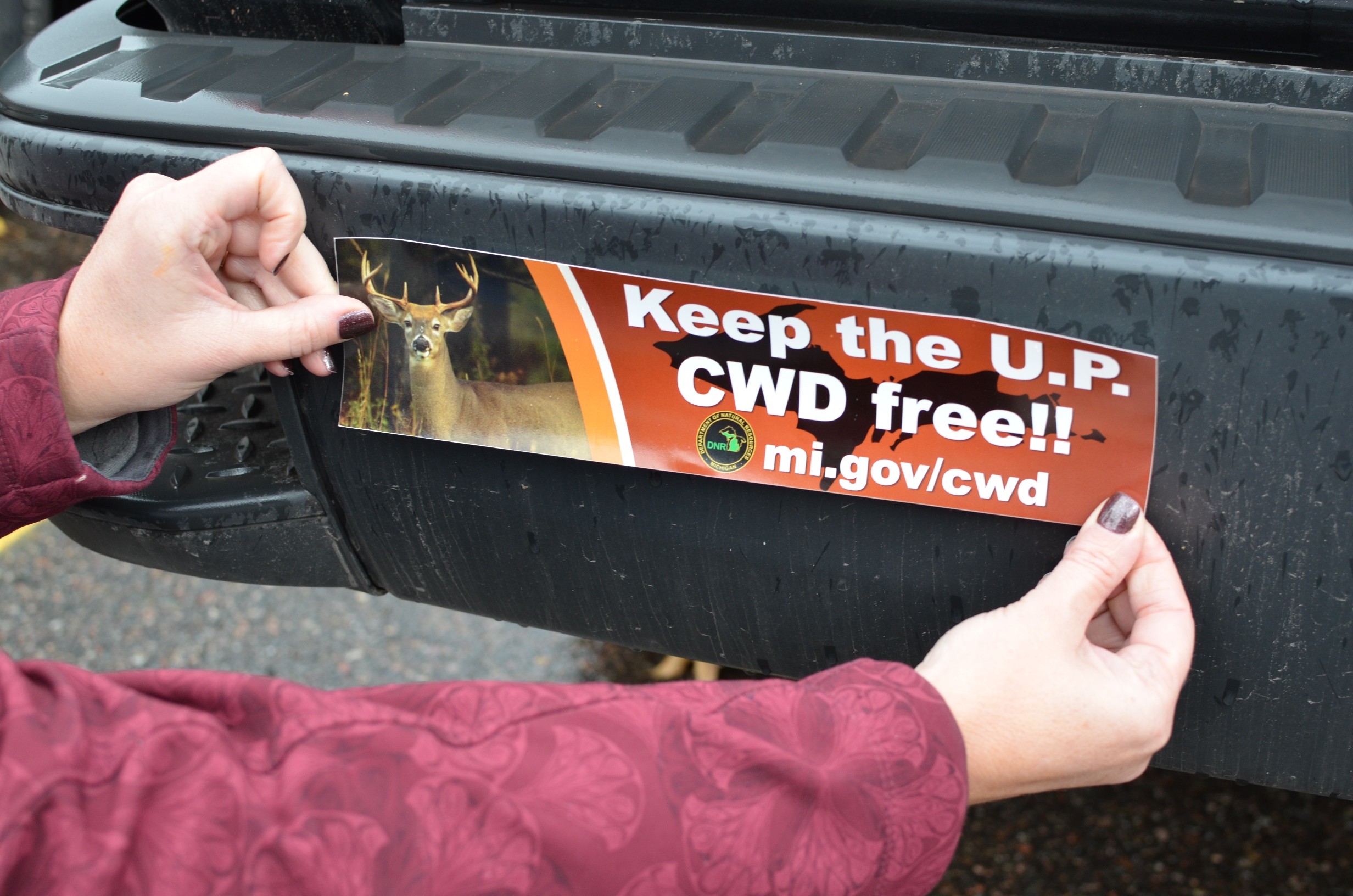